
Page 639 - Adams and Stashak's Lameness in Horses, 7th Edition
P. 639
Lameness of the Proximal Limb 605
VetBooks.ir
Figure 5.7. An adult horse with synovial effusion of the extensor
carpi radialis tendon sheath proximal to the carpal joints (arrows).
Source: Courtesy of Dr. Ty Wallis.
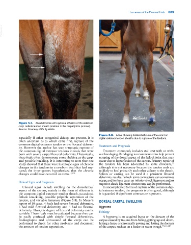
Figure 5.8. A foal showing bilateral effusion of the common
especially if other congenital defects are present. It is digital extensor tendon sheaths due to rupture of the tendons.
often uncertain as to which came first, rupture of the
common digital extensor tendon or the flexural deform- Treatment and Prognosis
ity. However the author has seen traumatic rupture of
the common digital extensor tendons in foals that were Treatment commonly includes stall rest with or with-
born with severe carpal flexural deformity. Historically, out bandaging. Bandaging is recommended to help protect
these foals often demonstrate some shaking at the carpi scraping of the dorsal aspect of the fetlock joint that may
and possible buckling. It is interesting to note that one occur due to hyperflexion of the carpus. Primary repair of
55
study showed that there were histologic signs of chronic the tendons has been advocated by some clinicians,
changes in the tendons in a newborn foal that had rup- although it is not necessary because the tendon ends are
tured; the investigators hypothesized that the chronic unlikely to heal primarily and rather adhere to the sheath.
changes could have occurred in utero. 13,132 Splints or casting can be used if a persistent flexural
deformity results. Fetlock joint contracture can sometimes
occur, and in these cases an inferior check ligament and/or
Clinical Signs and Diagnosis superior check ligament desmotomy can be performed.
Clinical signs include swelling on the dorsolateral In uncomplicated forms of rupture of the common digi-
aspect of the carpus, mainly in the form of effusion in tal extensor tendon, the prognosis is often good, although
the common digital extensor tendon sheath, occasional it is guarded if significant contracture is present.
fetlock knuckling, possible palpable separation of the
tendon, and variable lameness (Figure 5.8). In Meyer’s DORSAL CARPAL SWELLING
report of 10 cases, 4 foals had severe flexural deformity,
3 had mild flexural deformity, and 3 had no flexural Hygroma
deformity. Thus, the degree of flexural deformity can be Etiology
variable. These foals must be palpated because they can
be easily confused with simple flexural deformities. A hygroma is an acquired bursa on the dorsum of the
Radiographs and ultrasound of the carpi can be carpus caused by trauma from falling, getting up and down,
performed to check for other problems and document hitting a fence, or chronically pawing and hitting the dorsum
the amount of tendon separation. of the carpus, such as on a feeder or water trough. 99,115,119