
Page 644 - Adams and Stashak's Lameness in Horses, 7th Edition
P. 644
610 Chapter 5
VetBooks.ir
A B
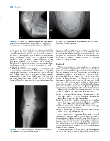
Figure 5.12. Radiographs demonstrating the need for a skyline any evidence of the fracture on the flexed lateral view (A). Source:
projection to characterize a slab fracture of the third carpal bone. Courtesy of Dr. Ryan Carpenter.
The fracture can be clearly seen on the skyline view (B) without
In the study by Simon and Dyson, lameness related to its cause and consequences are unknown. Ultimately,
the carpal joints was associated with increased ratio of arthroscopic surgery is usually the best diagnostic tool
radiopharmaceutical uptake and increased radiophar- to characterize and treat the problem in the carpus. The
maceutical uptake grade, both of which correlated with exception to this may be cases in which subchondral
opacity of the third carpal bone. In addition, intense bone pain is a primary problem without the overlying
108
uptake has been observed in 2‐year‐old Quarter horses articular cartilage damage.
that were worked on a treadmill for 6 months.
59
Therefore, it is important not to overinterpret the find- Treatment
ings because uptake can occur in horses that are under-
going active exercise. While some clinicians recommend rest for treatment
MRI and computed tomography have also been used of osteochondral fragmentation of the carpus, most sur-
to characterize subtle lesions in the carpus. Computed geons believe that arthroscopic removal of the fragments
tomography has higher resolution and can be used to and the resultant reduction of synovitis induced by the
detect both subtle lesions and bone mineral density instability provide a more predictable outcome. Most
abnormalities (Figure 5.14). MRI is useful for detecting surgeons feel that in horses that are conservatively
soft tissue damage and bone edema. Edema in the sub- treated, if these fragments are allowed to heal in an
90
chondral bone has been seen in joints with damage, but incongruent fashion, the fragment of bone can break
away later with training, again leading to clinical signs.
Arthroscopic surgery is the best method to fully charac-
terize the disease process, treat the primary problem,
and give an accurate prognosis for return to athletic use.
Degeneration of articular cartilage and bone has been
graded for severity and correlated with outcome: 81
Grade 1: Minimal fibrillation or fragmentation at the
edge of the defect left by the fragment, extending no
more than 5 mm from the fracture line
Grade 2: Articular cartilage degeneration extending
more than 5 mm back from the defect and including
up to 30% of the articular surface of that bone
Grade 3: Loss of 50% or more of the articular cartilage
from the affected carpal bone
Grade 4: Significant loss of subchondral bone (usually
distal radial carpal bone lesions)
After osteochondral fragments have been removed,
augmentative therapy such as microfracture or various
intra‐articular medications can be used if the lesions are
severe. There has been concern about operating horses
after they have been recently injected with corticoster-
oids because the horses may be predisposed to postop-
erative synovial sepsis; however, this has not been shown
to be of major concern.
Figure 5.13. A lateral radiograph of the carpus demonstrating a Arthroscopic surgery of the carpus is typically performed
slab fracture of the radial carpal bone (arrow). to remove the fragmentation or repair the fracture. Wright