
Page 647 - Adams and Stashak's Lameness in Horses, 7th Edition
P. 647
Lameness of the Proximal Limb 613
VetBooks.ir
A B
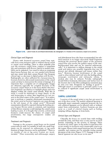
Figure 5.16. Lateromedial (A) and flexed lateromedial (B) radiographs of a fracture of the accessory carpal bone (arrows).
Clinical Signs and Diagnosis and debridement have also been recommended, but sub-
total resection is no longer advocated. Small fragments
Horses with fractured accessory carpal bone typi- involving the proximal dorsal aspect of the accessory
cally have acute lameness with or without dorsal carpal carpal bone may involve the palmar aspect of the ante-
or carpal canal swelling. There is often palpable pain brachiocarpal joint and can be removed arthroscopi-
over the accessory carpal bone, crepitus is sometimes cally. It is important to address damage to the carpal
62
felt, and sometimes with flexion, lateral to medial insta- canal and remove any possible fragments that may be
bility can be palpated. Care must be taken in examining present and debride damaged soft tissues within that
these horses because they are very painful with flexion area. However, because involvement of the carpal
85
and may stand with their carpus flexed. One frequent canal is often delayed until well into the healing phase,
13
clinical sign is a decrease in digital pulses with flexion. tenoscopic examination and retinacular release, if
Radiographs are often sufficient for characterizing the needed, also may be delayed. Periodic radiographs
fracture; however, ultrasound examination of the carpal should follow treatment; distraction and further lysis of
canal can help to characterize the extent of damage. It is the fracture due to the fibrocartilaginous nonunion may
important that a complete set of radiographic images be seen over time. Prognosis for healing is good. 62,85
are obtained, especially for complete fractures of the Although return to soundness is generally good, it can
accessory carpal bones as it has been shown that frac- be reduced if significant carpal canal involvement is
ture fragments can displace in multiple planes and even evident.
displace within the carpal canal distally. With this in
mind, it would be important to image distally to the
mid metacarpus to ensure that fragments are not pre- CARPAL LUXATIONS
sent in the distal aspect of the carpal canal. It is impor-
85
tant to perform a complete ultrasound examination of Luxation of the carpal joints is rare but can occur in
the carpal canal as fracture fragments can cause damage any of the three joints. The medial collateral ligament is
to the soft tissues of the carpal canal. Ultrasound reportedly most commonly ruptured; however, the lat-
85
examination should be repeated during the healing pro- eral collateral ligament can be affected with or without
cess because fibrous union is common, and expansion carpal bone comminution. Avulsion fractures can also
of this fibrous response could impinge the carpal canal, occur as a result of external trauma such as foaling,
leading to pain. It is thought that severely comminuted jumping, falling, slipping, or from kicks.
fractures could increase the chance of carpal canal
involvement, but large retrospective studies are
lacking. Clinical Signs and Diagnosis
Clinically, the horses are acutely lame with swelling
Treatment and Prognosis around the joint, and there may or may not be an ALD
present, depending on the severity of damage. These
Damage to the accessory carpal bone can be treated luxations are rarely open; in the author’s experience this
conservatively with 3–6 months of stall rest, small frag- occurs mostly in racehorses with catastrophic injuries to
ments can be removed via arthroscopy, or lag screw the carpus. Horses with carpal luxations may be axially
fixation of larger fractures can be performed. Three to unstable, and crepitus may be palpable during manual
31
six months of rest or lag screw fixation are recom- deviation of the joint (Figure 5.17). Damage to the col-
mended. Ulnar neurectomy and arthroscopic surgery lateral ligament can also occur on its own. 30
31