
Page 645 - Adams and Stashak's Lameness in Horses, 7th Edition
P. 645
Lameness of the Proximal Limb 611
VetBooks.ir
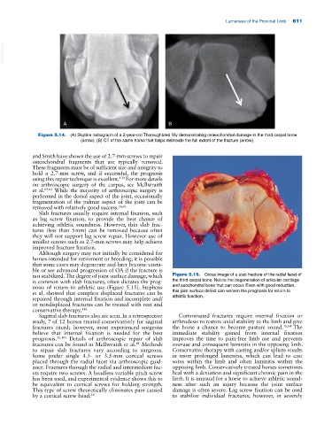
A B
Figure 5.14. (A) Skyline radiograph of a 2‐year‐old Thoroughbred filly demonstrating osteochondral damage in the third carpal bone
(arrow). (B) CT of the same horse that helps delineate the full extent of the fracture (arrow).
and Smith have shown the use of 2.7‐mm screws to repair
osteochondral fragments that are typically removed.
These fragments must be of sufficient size and integrity to
hold a 2.7‐mm screw, and if successful, the prognosis
using this repair technique is excellent. For more details
130
on arthroscopic surgery of the carpus, see McIlwraith
et al. 69,81 While the majority of arthroscopic surgery is
performed in the dorsal aspect of the joint, occasionally
fragmentation of the palmar aspect of the joint can be
removed with relatively good success. 26,62
Slab fractures usually require internal fixation, such
as lag screw fixation, to provide the best chance of
achieving athletic soundness. However, thin slab frac-
tures (less than 5 mm) can be removed because often
they will not support lag screw repair. However use of
smaller screws such as 2.7‐mm screws may help achieve
improved fracture fixation.
Although surgery may not initially be considered for
horses intended for retirement or breeding, it is possible
that some cases may degenerate and later become unsta-
ble or see advanced progression of OA if the fracture is
not stabilized. The degree of joint surface damage, which Figure 5.15. Gross image of a slab fracture of the radial facet of
is common with slab fractures, often dictates the prog- the third carpal bone. Notice the degeneration of articular cartilage
nosis of return to athletic use (Figure 5.15). Stephens and subchondral bone that can occur. Even with good reduction,
et al. showed that complete displaced fractures can be this joint surface defect can worsen the prognosis for return to
repaired through internal fixation and incomplete and/ athletic function.
or nondisplaced fractures can be treated with rest and
conservative therapy. 116
Sagittal slab fractures also are seen. In a retrospective Comminuted fractures require internal fixation or
study, 7 of 12 horses treated conservatively for sagittal arthrodesis to restore axial stability to the limb and give
fractures raced; however, most experienced surgeons the horse a chance to become pasture sound. 16,64 The
believe that internal fixation is needed for the best immediate stability gained from internal fixation
prognosis. 36,103 Details of arthroscopic repair of slab improves the time to pain‐free limb use and prevents
81
fractures can be found in McIlwraith et al. Methods overuse and consequent laminitis in the opposing limb.
to repair slab fractures vary according to surgeons. Conservative therapy with casting and/or splints results
Some prefer single 4.5‐ or 3.5‐mm cortical screws in more prolonged lameness, which can lead to cast
placed through the radial facet via arthroscopic guid- sores within the limb and often laminitis within the
ance. Fractures through the radial and intermediate fac- opposing limb. Conservatively treated horses sometimes
ets require two screws. A headless variable pitch screw heal with a deviation and significant chronic pain in the
has been used, and experimental evidence shows this to limb. It is unusual for a horse to achieve athletic sound-
be equivalent to cortical screws for holding strength. ness after such an injury because the joint surface
This type of screw theoretically eliminates pain caused damage is often severe. Lag screw fixation can be used
by a cortical screw head. 20 to stabilize individual fractures; however, in severely