
Page 648 - Adams and Stashak's Lameness in Horses, 7th Edition
P. 648
614 Chapter 5
VetBooks.ir
A B
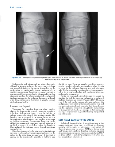
Figure 5.17. Radiographic images showing biaxial splint bone fractures (A, arrow) that led to instability and luxation of the carpus (B).
Source: Courtesy of Dr. Gary Baxter.
Radiographs and ultrasound are often diagnostic, should be used. Foals are usually casted for approxi-
although in some partial tears the horse must be sedated mately 4 weeks and adults for 6 weeks to allow fibrosis
and manual deviation of the carpus imposed to see the to occur in the collateral ligament area and joint cap-
subluxation on radiographs (stress radiographs). In sule. The horse may be transferred to a bandage and/or
addition, dorsopalmar luxations can occur and radio- splint for several weeks, but stall confinement for sev-
graphs should be assessed closely. Ultrasound is the best eral months is necessary.
diagnostic method for characterizing full‐ and partial‐ Partial or pancarpal arthrodesis may be needed to
thickness damage to the collateral ligaments, although stabilize the limb for breeding and pasture soundness if
22
with time, enthesophyte formation is usually appreci- multiple fractures are present (Figure 5.18). However,
ated radiographically. even if the limb can be reduced adequately, chronic OA
and pain may necessitate arthrodesis to control pain and
Treatment and Prognosis prevent contralateral limb laminitis. Soft tissue lesions
can be augmented with stem cells, platelet‐rich plasma,
Treatment for complete luxations often involves or extracorporeal shockwave therapy to help stimulate
placing the horse under general anesthesia to achieve healing. The prognosis is good for healing but guarded
reduction. Arthroscopic surgery may be helpful to for athletic use.
debride damaged tissues if joint damage occurs. The
luxation can be reduced if the carpal bones are not
involved; otherwise, fractures may need to be debrided SOFT TISSUE DAMAGE TO THE CARPUS
to facilitate reduction. Dorsopalmar luxations can be
difficult to reduce, and sometimes manual fatiguing of Collateral ligament injury is sometimes seen in the
the limb or surgery is needed to reduce the luxations. carpus. Although these may be difficult to diagnose
Once reduced, the limb can be put through rotational acutely, improvement in ultrasound examination of
manipulation. these structures and the use of MRI have helped with
If the limb is deemed to be rotationally stable, then a early diagnosis. Some have described collateral ligament
tube cast can be applied from the proximal aspect of the abnormalities in the carpus closely associated with OA;
30
radius to the distal third metacarpus. If the limb is so the problem is sometimes diagnosed well after OA
13
deemed to be rotationally unstable, a full‐limb cast has already been established.