
Page 652 - Adams and Stashak's Lameness in Horses, 7th Edition
P. 652
618 Chapter 5
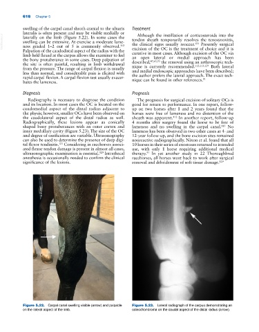
swelling of the carpal canal sheath cranial to the ulnaris Treatment
lateralis is often present and may be visible medially or Although the instillation of corticosteroids into the
VetBooks.ir swelling can be extensive. At exercise a moderate lame- tendon sheath temporarily resolves the tenosynovitis,
laterally on the limb (Figure 5.22). In some cases the
the clinical signs usually reoccur. Presently surgical
121
ness graded 1–2 out of 5 is commonly observed.
112
Palpation of the caudodistal aspect of the radius with the excision of the OC is the treatment of choice and it is
limb held flexed at the carpus allows the examiner to feel curative in most cases. Although excision of the OC via
the bony protuberance in some cases. Deep palpation of an open lateral or medial approach has been
47,63,70
the site is often painful, resulting in limb withdrawal described, the removal using an arthroscopic tech-
112,113,129
from the pressure. The range of carpal flexion is usually nique is currently recommended. Both lateral
less than normal, and considerable pain is elicited with and medial endoscopic approaches have been described;
rapid carpal flexion. A carpal flexion test usually exacer- the author prefers the lateral approach. The exact tech-
81
bates the lameness. nique can be found in other references.
Diagnosis Prognosis
Radiography is necessary to diagnose the condition The prognosis for surgical excision of solitary OCs is
and its location. In most cases the OC is located on the good for return to performance. In one report, follow‐
caudomedial aspect of the distal radius adjacent to up on two horses after 1 and 2 years found that the
the physis; however, smaller OCs have been observed on horses were free of lameness and no distention of the
the caudolateral aspect of the distal radius as well. sheath was apparent. 112 In another report, follow‐up
Radiographically, these lesions appear as conically 4 months after surgery found the horse to be free of
shaped bony protuberances with an outer cortex and lameness and no swelling in the carpal canal. No
113
inner medullary cavity (Figure 5.23). The size of the OC lameness has been observed in two other cases at 4‐ and
and degree of ossification are variable. Ultrasonography 12‐year follow‐up, and the bone excision sites remained
can also be used to determine the presence of deep digi- nonreactive radiographically. Nixon et al. found that all
tal flexor tendinitis. Considering in racehorses associ- 10 horses in their series of exostoses returned to intended
121
ated flexor tendon damage is present in almost all cases, use, with only 1 horse requiring additional medical
ultrasonographic examination is essential. Intrathecal therapy. In yet another study in 22 Thoroughbred
129
91
anesthesia is occasionally needed to confirm the clinical racehorses, all horses went back to work after surgical
significance of the lesions. removal and debridement of soft tissue damage. 129
Figure 5.22. Carpal canal swelling visible (arrow) and palpable Figure 5.23. Lateral radiograph of the carpus demonstrating an
on the lateral aspect of the limb. osteochondroma on the caudal aspect of the distal radius (arrow).