
Page 651 - Adams and Stashak's Lameness in Horses, 7th Edition
P. 651
Lameness of the Proximal Limb 617
OSTEOCHONDROSIS OF THE CARPUS few other long bones, rather than numerous growing
bones. Solitary OCs have been reported in two foals; the
VetBooks.ir carpal bones, although it is uncommon compared to in one, and in the other the calcaneus was involved. 24,32
Osteochondritis dissecans has been reported in the
distal palmar aspect of the middle phalanx was affected
other joints in the horse. Subchondral cystic lesions in
The reader is referred to Chapter 7 for more details.
some carpal bones are incidental and are often of ques-
tionable clinical significance. This is especially true with
cysts within the second carpal bone in association with Etiology
the presence of the first carpal bone and in the proximal Although a single dominant autosomal gene is
aspect of the second metacarpal bone. However, sub- responsible for the development of multiple exostosis in
chondral cystic lesions in the radiocarpal bone and the humans and horses, the genetic implications for iso-
107
distal aspect of the radius (Figure 5.21) are often clini- lated OC remains unclear. In humans, solitary OCs are
cally significant, and arthroscopic surgery and debride- not considered to be inherited. It is postulated that
51
ment are warranted if they are felt to communicate with metaplastic cartilage foci develop in the metaphysis and
the carpal joints. Simple drilling or transcystic screw distal diaphysis from abnormal growth of the perios-
could also be considered in these cases. Oftentimes, MRI teum. As the cartilage grows, it undergoes endochondral
or CT is needed to best characterize these lesions. ossification similar to that occurring at the physis. The
developing exostosis, which is continuous with the cor-
OSTEOCHONDROMA OF THE DISTAL RADIUS tex of the bone and surrounded by a cartilage cap, is
called an osteochondroma.
Osteochondroma (OC) formation at the distal end of Nixon et al. have reported on a series of cases in
the radius is an uncommon condition causing lameness which exostoses of the palmar aspect of the closed
in the horse. 53,63,70,114 The growth most commonly occurs physis were removed arthroscopically. In these cases,
in adult horses adjacent to the physis at the caudal distal although they appeared similar to OC, they were histo-
aspect of the radius. 47,112,113 Although very uncommon, logically different. 91
the cranial aspect of the radius also can be involved.
104
Radiographically and histologically, these new bone Clinical Signs
growths appear much like those reported for hereditary
multiple exostosis. However, unlike hereditary multiple Affected horses often present with a history of inter-
exostosis, they appear as singular lesions or affect only a mittent lameness that increases with exercise. An obvious
Figure 5.20. A DP radiograph demonstrating collapse of the
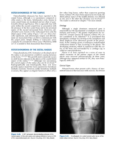
medial aspect of the joint space and osteoproliferation typical of OA Figure 5.21. A radiograph of a subchondral cystic lesion of the
of the carpometacarpal joint. Source: Courtesy of Dr. Gary Baxter. distal radius (arrow). Source: Courtesy of Dr. Gary Baxter.