
Page 640 - Adams and Stashak's Lameness in Horses, 7th Edition
P. 640
606 Chapter 5
Clinical Signs and Diagnosis mass with a Penrose drain and bandaging has been used
2
successfully for treatment of recurrent hygromas.
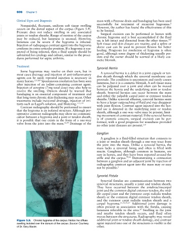
Nonpainful, fluctuant, uniform soft tissue swelling
VetBooks.ir occurs on the dorsal aspect of the carpus (Figure 5.9). However, the author has found the effectiveness of this
to be limited.
Pressure does not induce swelling in any associated
Surgical excision can be performed in horses with
joints or tendon sheaths. Range of motion of the carpus
may be reduced, but lameness is unusual. However, chronic hygroma and is best accomplished if the fluid
sac is left intact and dissected from the other tissues.
113
lameness can be severe if the hygroma is infected.
Injection of radiopaque contrast agent into the hygroma Soft tissue and skin closure are routine, and a splint or
sleeve cast can be used to prevent flexion for better
confirms its extra‐articular position. If a hygroma is sus-
pected of being infected, then a fluid sample should be healing. Prognosis for resolution of hygroma is often
good, although some degree of thickening usually per-
submitted for cytology and culture, similar to the proce-
dures performed for septic arthritis. sists and the owner should be warned of a likely cos-
metic blemish.
Treatment Synovial Hernia
Some hygromas may resolve on their own, but in
most cases drainage and injection of anti‐inflammatory A synovial hernia is a defect in a joint capsule or ten-
agents can be used; repeated injection is necessary in don sheath through which the synovial membrane can
protrude. The condition is uncommon and rarely causes
many horses. 115,119 Spontaneous resolution has been seen
after injection of an iodine‐containing contrast agent. lameness, but it is a cosmetic blemish. A soft tissue mass
can be palpated over a joint and often can be moved
Injection of atropine (7 mg total dose) may also help to
resolve the swelling. Owners should be warned that between the hernia and the underlying joint or tendon
sheath. Synovial hernias can occur between the mass
bandaging is an essential component of treatment and
that long‐term chronic skin thickening may occur. Other and either the palpable carpal joint or extensor tendon
sheaths. Unlike synovial effusion, synovial hernias seem
treatments include incisional drainage, injection of irri-
tants such as Lugol’s solution, and blistering. 115,119 to have a larger outpouching of fluid and may disappear
with joint flexion. Contrast agent injected into the her-
Contrast radiography should be performed to ensure
that the hygroma is an isolated structure. Although pre- nial sac is detected in the underlying joint or tendon
sheath, although a one‐way valve may be present, limit-
operative contrast radiographs may show no communi-
cation between a hygroma and a joint or tendon sheath, ing movement of contrast material. If the synovial hernia
is of cosmetic concern, surgical excision can be per-
it is possible that one exists in the form of a one‐way
valve from the joint into the mass. 52,99 Drainage of the formed, with a good prognosis for soundness provided
no other joint diseases are present. 113
Ganglion
A ganglion is a fluid‐filled structure that connects to
a joint or tendon sheath through a one‐way tract from
the joint into the mass. Unlike a synovial hernia, the
mass lacks a synovial lining and often is filled with
mucin. Ganglions, although common in humans, are
rare in horses, and they have been reported around the
stifle and the carpus. 75,99 Demonstrating a connection
between a ganglion and an adjacent joint by injection of
radiographic contrast agent into the mass may or may
not be possible. 2
Synovial Fistula
Synovial fistulae are communications between two
synovial structures, usually a joint and tendon sheath.
They have occurred between the antebrachiocarpal
joint and the common digital extensor tendon, the mid-
dle carpal joint and the extensor carpi radialis tendon
sheath or the common digital extensor tendon sheath,
and the extensor carpi radialis tendon sheath and a
carpal hygroma. 21,52,56,67 Additional joint damage is
often present in association with the fistula, causing
lameness referable to the area. Swelling in the joint
67
and nearby tendon sheath occurs, and fluid often
moves between the structures. Radiography may reveal
Figure 5.9. Chronic hygroma of the carpus. Notice the diffuse additional joint or tendon sheath damage, and contrast
swelling isolated over the dorsum of the carpus. Source: Courtesy agent injected into one of the structures is visible in the
of Dr. Gary Baxter. other.