
Page 754 - Adams and Stashak's Lameness in Horses, 7th Edition
P. 754
720 Chapter 5
VetBooks.ir
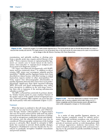
Figure 5.124. Ultrasound images of a middle patellar ligament injury. The cross‐sectional view on the left demonstrates two areas of
reduced fiber density (#2 and #3). The black arrows on the longitudinal view on the right outline the same areas at the same horizontal level
as the image on the left. Source: Courtesy of Dr. Anne Desrochers.
examination, and palpable swelling or eliciting pain
from a specific point may require partial flexion of the
stifle. More extensive lesions or ruptured patellar liga-
28
ments are usually easy to locate based on the horse’s
stance. Intra‐articular or regional local analgesia pro-
duces equivocal diagnostic results. 28
LPL injury was observed simultaneously with MidPL
injury and less commonly alone. In addition to direct
28
trauma, MPL injury may accompany palpable patellar
instability. Middle patellar ligament lesions have been
28
described as a linear lucency with fiber tearing or central
hypoechoic regions similar to other desmopathies. 28,107
Some suspect that ultrasonographic abnormalities in
patellar ligaments detected after injury bear more patho-
physiologic significance than those reported following
MPD. Proximal and distal enthesopathies may reflect
bony disruption in addition to the soft tissue lesion,
79
but their role in lameness if the inciting inflammation
subsides is unclear. 80,107
The MPL is affected less commonly by primary desmi-
tis. Similar trauma seems to cause avulsion fracture of
the medial aspect of the patella instead of ligament inju-
ries. However primary MPL rupture can occur but usu- Figure 5.125. This horse developed a ruptured medial patellar
9
ally heals quickly with stall confinement (Figure 5.125). ligament with an obvious palpable defect after hitting a jump. It
healed completely and the horse became sound, although there
were subtle radiographic changes on the distal patella.
Treatment
When the injury is confined to the soft tissue, therapy
consists of rest and local and systemic anti‐inflamma-
tory therapy. Smaller enthesopathies or fragmentation
also may be treated conservatively with stall rest and/or Prognosis
extracorporeal shockwave therapy. Injections of biolog- In a series of nine patellar ligament injuries, no
ics, such as autogenous conditioned serum and platelet‐ horses became completely sound for athletic perfor-
rich plasma, into the affected ligament(s) may also be mance, although working soundness was achieved in
considered. Ultrasonographic monitoring of the healing one. These injuries may heal better with prolonged
28
process is advisable. Rest should be extended if MPL periods of restricted activity or with desmoplasty with
laxity has occurred to prevent distal patellar changes. or without regenerative medicine intervention. Pain
Progressive rehabilitation exercises should also be con- control should be used cautiously with continued train-
sidered to gradually strengthen hindlimb musculature ing due to the potential for patella instability leading to
and provide mobility. secondary OA.