
Page 749 - Adams and Stashak's Lameness in Horses, 7th Edition
P. 749
Lameness of the Proximal Limb 715
may be more efficient to use the direct approach. The
prognosis for these horses is usually reasonable for nor-
VetBooks.ir Intra‐articular Fractures: Patella
mal activity.
Patellar fractures occur infrequently in horses, but
several configurations including sagittal, 3,23,27,59 trans-
verse, comminuted, basilar (proximal), 18,27,108 and
47
77
distal fragmentation of the patella 62,84 have been
described. Combinations of types of patellar fracture
and associated fracture of the femur may also occur. 27
Etiology
Direct trauma to the patella while the stifle joint is in
a semiflexed position is commonly the cause. The patella
is immobilized when the stifle is semiflexed, making it
more susceptible to direct trauma. Horses that jump
21
can strike jumps and sustain bilateral patellar frac-
tures, or the fractures can occur as a result of a kick.
59
Sudden lateral slips may cause a separation of the medial
fibrocartilage. The prominence of the medial trochlear
23
ridge may be a point of contact, causing a relative higher
incidence of fractures toward the medial side of the
patella.
Some horses suffer fragmentation of the distal patella
(FDP) when they return to work too soon after undergo-
ing a medial patellar ligament desmotomy (MPD) and
may be caused by direct trauma secondary to temporary
patellar instability. 62,84 This condition is discussed in the
next section.
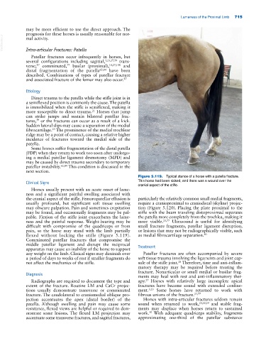
Figure 5.119. Typical stance of a horse with a patella fracture.
Clinical Signs This horse had been kicked, and there was a wound over the
cranial aspect of the stifle.
Horses usually present with an acute onset of lame-
ness and a significant painful swelling associated with
the cranial aspect of the stifle. Femoropatellar effusion is particularly the relatively common small medial fragments,
usually profound, but significant soft tissue swelling require a cranioproximal to craniodistal (skyline) projec-
may obscure palpation. Pain and sometimes crepitation tion (Figure 5.120). Placing the plate proximal to the
may be found, and occasionally fragments may be pal- stifle with the beam traveling distoproximad separates
pable. Flexion of the stifle joint exacerbates the lame- the patella more completely from the trochlea, making it
ness and the painful response. Weight‐bearing may be more visible. 23,77 Ultrasound is useful for identifying
difficult with compromise of the quadriceps or from small fracture fragments, patellar ligament disruption,
pain, so the horse may stand with the limb partially or lesions that may not be radiographically visible, such
flexed without locking the stifle (Figure 5.119). as medial fibrocartilage separation. 80
Comminuted patellar fractures that compromise the
middle patellar ligament and disrupt the reciprocal Treatment
apparatus may cause an inability of the horse to support
any weight on the limb. Clinical signs may diminish over Patellar fractures are often accompanied by severe
a period of days to weeks of rest if smaller fragments do soft tissue trauma involving the ligaments and joint cap-
not affect the mechanics of the stifle. sule of the stifle joint. Therefore, time and anti‐inflam-
28
matory therapy may be required before treating the
fracture. Nonarticular or small medial or basilar frag-
Diagnosis
ments may heal with rest and anti‐inflammatory ther-
27
Radiographs are required to document the type and apy. Horses with relatively large incomplete apical
extent of the fracture. Routine LM and CaCr projec- fractures have become sound with extended confine-
tions usually demonstrate transverse or comminuted ment. 3,23 Some horses have returned to work with
fracture. The caudolateral to craniomedial oblique pro- fibrous unions of the fracture. 23,77
jection accentuates the apex (distal border) of the Horses with intra‐articular fractures seldom remain
patella. Although swelling and pain may cause some sound when returned to work, 23,30,59 and stable frag-
resistance, flexed views are helpful or required to dem- ments may displace when horses return to sustained
onstrate some lesions. The flexed LM projection may work. With adequate quadriceps stability, fragments
30
accentuate some transverse fractures, and sagittal fractures, approximating one‐third of the patellar substance