
Page 747 - Adams and Stashak's Lameness in Horses, 7th Edition
P. 747
Lameness of the Proximal Limb 713
In summary, a minority of horses with mild lateral for systemic treatment. Intra‐articular corticosteroids
15
trochlear ridge defects have no cartilage defect during are inadvisable where cartilage healing is needed, and
VetBooks.ir severe radiographic changes probably have cartilage ture equine joints. The excess fluid can be drained at
surgery (Figure 5.116). Conversely, horses with more
negative effects of steroids have been observed in imma-
39
defects at that location and possibly elsewhere in the
the time of treatment so that resolution of the synovitis
same joint. When radiographic lesions are absent, lame- can be monitored. In one study, half of 23 affected foals
ness and synovial effusion often dictate the clinical sig- that were diagnosed early with OCD and treated with
nificance of the OCD lesion. stall rest eventually raced. Generally, the successfully
66
treated horses tended to have less severe lesions.
Sequential radiographs and clinical response to ther-
Treatment
apy are used to determine whether surgery is advisable.
Surgical management is the mainstay of treatment for Surgery has been recommended for young horses with
OC in young athletic horses. Surgical debridement is radiographic lesions greater than 2 cm in length or
76
also thought to produce better results than conservative deeper than 5 mm or lesions with ossification in the
therapy when clinical signs are present in adults. defect and femoropatellar effusion. 63,65 However, the
63
However, in one study, some lesions detected up to 8 need to progress with the horse’s conditioning schedule
months of age were found to resolve over time, and no may dictate earlier surgical intervention, or preemptive
24
worsening of the lesion occurred after 11 months of age. removal is often requested by some owners and trainers
Stall confinement is recommended in very young horses in order to minimize the chances of the lesion affecting
with OCD lesions to protect the articular surface from training and early competition.
disruption and to facilitate healing, but surgery is indi- Weanlings and short yearlings can present with effu-
66
cated if the clinical signs persist after 1 year of age. 63,76 sion and lameness but no radiographic lesions. 9,66
Intra‐articular and systemic anti‐inflammatory ther- Conservative therapy is indicated unless clinical signs
apy to minimize the chance of degenerative change and persist. Subsequent radiographs may demonstrate heal-
improve cartilage healing is advisable in young horses. ing or larger trochlear ridge ossification defects because
Biologic therapies such as autogenous conditioned the lesion has persisted while the surrounding trochlear
serum are recommended for intrarticular (IA) treatment, ridge has ossified normally as the epiphysis has
19
and polysulfated glycosaminoglycan is most often used expanded. Some believe that surgical lesions in foals
and weanlings may progress, suggesting that surgery
should be delayed until the lesion has fully developed.
19
This consideration will have passed after a period of
conservative therapy. Medical therapy in cases without
radiographic abnormalities can be diagnostic as well
because a permanent response indicates that there is
likely no articular surface lesion.
Arthroscopic surgery is indicated when it is obvious
that the lesion will not heal with conservative therapy
and in horses beyond a year of age with persistent
clinical signs. The technique for arthroscopic debride-
ment of femoropatellar OCD lesions has been well
described, 63,65,68,76,101 and there are multiple different
techniques and approaches. 65,68 The objective of surgery
is to remove all loose osteochondral tissue and debride
the lesion to leave healthy subchondral bone to fill with
fibrocartilage and resolve the synovitis (Figure 10.49
in Chapter 10). A thorough evaluation of the joint is
indicated as silent OCD lesions may be visible with the
arthroscope that were not apparent on radiographs. It is
also important to remove all the debris generated during
the debridement process because it can contribute to
continued synovitis. 48,68 Arthroscopic reattachment of
OCD cartilage flaps has been reported in select horses
but is not performed routinely. 90
Prognosis
The prognosis for athletic activity following arthro-
scopic surgery for femoropatellar OCD is generally
good. Of a series of 134 horses, only 16% were unsuc-
34
cessful for reasons related to the OCD. Slightly fewer
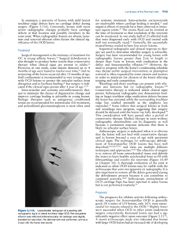
Figure 5.116. Lateromedial radiograph of a yearling with were successful when OCD in other joints underwent
radiographic signs of lateral trochlear ridge OCD. Femoropatellar surgery concurrently. Increased lesion size had a sig-
effusion was mild and arthroscopically the cartilage was slightly nificantly negative effect upon outcome (Figure 5.117).
elevated but was intact. No debridement was performed, and as a A recent arthroscopic study also indicated that horses
3‐year‐old, the horse was sound. with larger OCD lesions had an increased risk of developing