
Page 744 - Adams and Stashak's Lameness in Horses, 7th Edition
P. 744
710 Chapter 5
demonstrate a subtle lameness under saddle or during A complete series of good‐quality radiographs is the
specific movements. initial imaging tool to evaluate the stifle joints in most
VetBooks.ir and hip. Digital flexion is minimized if the limb is held obtain very good films in the field. Routine survey
cases. Digital radiography has made it possible to
Flexion of the hindlimb affects the digit, hock, stifle,
7
10
loosely around the fetlock. However, the source of any
views include caudocranial (CaCr) and lateromedial
increased pain following flexion must still be localized. (LM) views. The 30° lateral craniomedial oblique sepa-
The upper limb flexion (commonly referred to as the rates the lateral from the medial trochlear ridge, if that
35
stifle flexion test) can provide the examiner with some is needed. The articular surface and sagittal integrity of
assuredness that the region is involved if the horse reacts the patella are demonstrated best by a flexed cranio-
in a painful manner to flexion and the test exacerbates proximal to craniodistal tangential (skyline) view. 23,30,83
the lameness. See Chapter 2 for more information on Some horses may have radiographically inapparent
flexion tests. However, the stifle flexion test is not spe- lesions or more extensive bony involvement than radio-
cific for the stifle. graphs demonstrate. 19,67
Lameness can be localized to the stifle with intra- Ultrasonography is extremely helpful for evaluating
synovial anesthesia if necessary. In some cases in which the femoropatellar and femorotibial regions. The charac-
physical examination shows strong indications of sti- ter of the articular surfaces, menisci and subchondral
fle disease, diagnostic imaging may be performed prior bone, 11,22,78 joint capsule, synovial fluid, 60,79 patellar and
67
78
to intrasynovial anesthesia in order to prevent fluid or intra‐articular ligaments, and periarticular tissues can
air artifact from interfering with image quality. The be viewed with ultrasound. 22,67,79,80 However, the articular
number and sequence of blocking is dependent on the surface of the patella is not readily accessible. 7,67
signalment of the horse, clinical findings, and clinician The benefit of scintigraphy for diagnosis of stifle
preference. For instance, a western performance horse lameness is variable. Some have reported it beneficial for
with effusion of the MFT joint would warrant block- lesions in the stifle region, 67,71 whereas others have ques-
ing the MFT joint alone. A horse with lameness that tioned its ability to diagnose osteochondrosis (OC) or
you are trying to rule out the stifle as the location of subchondral cystic lesions due to the lack of increased
lameness may warrant blocking all three joints simul- vascularity or increased bone remodeling required to
taneously. Without effusion, the MFT joint is the logi- sequester the radiopharmaceutical. Normal horses
92
cal starting point if each joint is to be blocked have patterns of uptake highlighting the patella and cau-
separately, because the FP joint will usually have effu- doproximal tibia that should not be mistaken for patho-
sion if there is a problem, and the LFT joint is not the logic change, especially in young athletic horses. 31
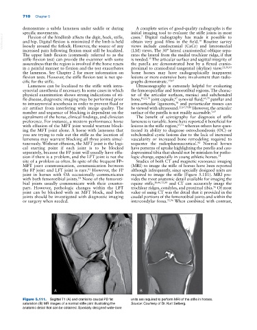
site of a problem as often. In spite of the frequent FP– Studies of both CT and magnetic resonance imaging
MFT joint communication, communication between (MRI) to image the stifle of horses have been reported
the FP joint and LFT joint is rare. However, the FP although infrequently, since specially designed units are
81
joint in horses with OA occasionally communicates required to image the stifle (Figure 5.111). MRI pro-
with both femorotibial joints. None of the femoroti- vides the most anatomic detail available for imaging the
81
bial joints usually communicate with their counter- equine stifle, 20,46,73,88 and CT can accurately image the
part. However, pathologic changes within the LFT trochlear ridges, condyles, and proximal tibia. Of most
96
joint can be blocked with an MFT block, and both value of using CT was the detail that it provided in the
joints should be investigated with diagnostic imaging caudal portions of the femorotibial joints and within the
or surgery when needed. intercondylar fossa. 72,96 When combined with contrast,
A B
Figure 5.111. Sagittal T1 (A) and cranial to caudal PD fat units are required to perform MRI of the stifle in horses.
saturation (B) MR images of a normal stifle joint illustrating the Source: Courtesy of Dr. Kurt Selberg.
anatomic detail that can be obtained. Specially designed wide‐bore