
Page 739 - Adams and Stashak's Lameness in Horses, 7th Edition
P. 739
Lameness of the Proximal Limb 705
Incomplete fissure fractures of the distal metaphysis chondrocytes within the physis and breaks out into the
of the tibia may benefit from a cast. These can be applied lateral metaphysis. This is a consistent configuration of
VetBooks.ir ing position. Stall rest is recommended until there is evi- to medial bending as the primary force responsible for
in the sedated standing horse in a normal weight‐bear-
proximal tibial physeal fractures and implicates lateral
the injury.
dence of fracture healing. This usually requires a
minimum of 3–4 months. Maintaining a cast on the Blunt trauma to the lateral aspect of the stifle while
hindlimb can be problematic and may require several the foal is weight bearing on the limb can create this
cast applications/resets to keep the horse comfortable type of physeal fracture. This injury can also occur when
on the limb. During the last 30 days of confinement, a recumbent foal attempts to stand while the uppermost
once the cast has been removed, a program that incor- limb is positioned under a stationary object or when the
porates a gradual increase in hand walking is recom- mare steps on the recumbent foal’s uppermost hindlimb.
mended. In the best of circumstances, these fractures can There is usually marked soft tissue swelling on the
heal without significant complications. However, propa- medial aspect of the physis. These fractures can be rela-
gation of the fracture is not uncommon. Careful daily tively straightforward to repair with a good prognosis
monitoring to assess the comfort level of the horse may for healing but not necessarily for soundness. Fracture
provide an indication about the integrity and stability of repair is best achieved by reestablishing support on the
the fracture. Any trend toward increasing discomfort medial aspect of the proximal tibia. Screw fixation, pin-
may indicate a compromise in the stability of the frac- ning, and plate fixation have all been described for treat-
ture and a need for surgical repair. ing these fractures either alone or in combination. 3,12,13,34,35
Medial plate application is currently the most common
PROXIMAL PHYSEAL FRACTURES fixation technique utilized for repair and can be signifi-
cantly improved by the placement of a tension band
The bones of young horses are more elastic than along the tibial crest. This neutralizes the distracting
mature animals and are less likely to develop into forces of the quadriceps muscle that is transmitted
severely comminuted fractures. Young horses have an across the stifle joint to the tibial crest via the patella
increased risk to develop fractures through the epiphy- and the patellar ligaments’ attachment to the tibial crest.
seal growth plate (physis). Physeal fractures are called These fractures usually heal quickly and the progno-
Salter–Harris (SH) fractures and are further classified sis is good. Rapid reestablishment of the vascular system
based on the relative involvement of the physis, meta- to the physis occurs with the stabilization of the fracture
physis, and epiphysis. 3,12,13,33 The young horse that devel- and allows fracture healing to occur quickly, normally in
ops a tibial fracture will most commonly develop an SH 3–4 weeks. The amount of damage to the soft tissues,
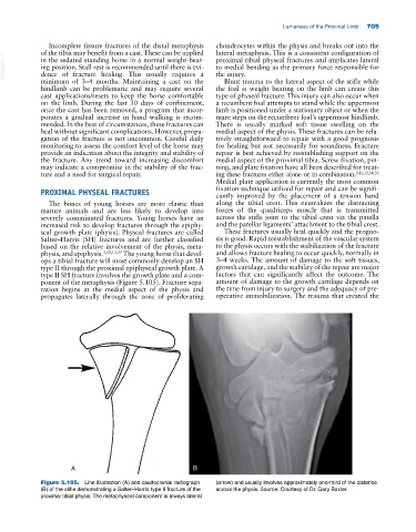
type II through the proximal epiphyseal growth plate. A growth cartilage, and the stability of the repair are major
type II SH fracture involves the growth plate and a com- factors that can significantly affect the outcome. The
ponent of the metaphysis (Figure 5.105). Fracture sepa- amount of damage to the growth cartilage depends on
ration begins at the medial aspect of the physis and the time from injury to surgery and the adequacy of pre-
propagates laterally through the zone of proliferating operative immobilization. The trauma that created the
A B
Figure 5.105. Line illustration (A) and caudocranial radiograph (arrow) and usually involves approximately one‐third of the distance
(B) of the stifle demonstrating a Salter–Harris type II fracture of the across the physis. Source: Courtesy of Dr. Gary Baxter.
proximal tibial physis. The metaphyseal component is always lateral