
Page 740 - Adams and Stashak's Lameness in Horses, 7th Edition
P. 740
706 Chapter 5
fracture often damages the physis leading to growth dis-
turbances. The entire growth plate can close, which can
VetBooks.ir growth in the rest of the limb may counteract the short-
lead to shortening of the tibia. Compensatory bone
ening of the tibia. Implant removal may be considered to
prevent or reduce the likelihood of premature physeal
closure and limb shortening. This can be done in most
instances at 8 weeks postoperatively. In general, the
smaller the foal, the better the prognosis.
DIAPHYSEAL FRACTURES
Fractures of the diaphysis of the tibia are the second
most common type of tibial fracture and occur in all
ages of horses. Fracture type can range from simple
oblique to highly comminuted. 3,6–9,13,15,16,26,29 Diaphyseal
fractures are thought to occur when the tibia is loaded
under considerable torque in combination with an
excessive external force. Most diaphyseal fractures have
a spiral configuration and many are severely commi-
nuted. These fractures are characterized by sudden onset
of non‐weight‐bearing lameness. Diaphyseal fractures
are easily diagnosed but require radiographs to assess
the fracture configuration. These fractures are often cat-
astrophic injuries characterized by extensive comminu-
tion especially in mature horses. However, simple long
oblique fractures of the mid‐diaphysis do occur and are
often repairable provided that they are not open. Because
the tibia has very little muscle coverage on the medial
aspect, fracture fragments often disrupt the skin and soft
tissues (Figure 5.101). Attempts at repair of open tibial
fractures are seldom practical because of the very poor
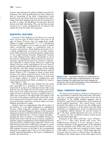
prognosis. However, diaphyseal fractures in foals tend Figure 5.106. Lateromedial radiograph of a tibial fracture in a
to be simple oblique or spiral in configuration and foal at the time of repair using a single plate placed on the cranial
closed, making foals much better candidates for surgical aspect of the tibia. The single screw was used to compress the
repair (Figure 5.106). fracture line. Note the solid caudal buttress on the caudal aspect of
The Thomas splint has been utilized but has rarely the tibia. Source: Courtesy of Dr. NA White.
been successful as a treatment for tibial fractures in
some foals. It consists of an aluminum frame with a ring
that fits over the thigh and into the groin of the horse TIBIAL TUBEROSITY FRACTURES
with cranial and caudal extensions reaching the foot to
apply traction to the limb. Traction is applied with 1‐in. The large cranial eminence of the proximal aspect of
white tape attached to the distal limb and pulled to the the cranial tibia is called the tibial tuberosity. This tuber-
lower bar of the splint to stabilize the entire limb. Long‐ osity has a prominent groove that serves as the site for
term management of fractures of the tibia in this manner attachment of the middle patellar ligament and is
can become quite expensive, and these splints are often flanked by rough areas for attachment of the medial and
difficult and tedious to apply. lateral patellar ligaments. The cranial border of the
14
Surgical repair is compromised when the location of proximal tibia distal to the groove for the middle patel-
the fracture is in the distal diaphyseal or metaphyseal lar ligament is referred to as the tibial crest. The tibial
location, which frequently precludes adequate implant tuberosity is a supplementary center of ossification that
purchase in the distal fragment. Because of the reciprocal is completely ossified between 36 and 42 months of
apparatus, traction on the distal limb does not help reduce age. The three patellar ligaments also serve as the site
14
a diaphyseal tibial fracture—in fact, it causes overriding— of insertion for the quadriceps femoris muscle group
hanging the limb in tension is not helpful. Placing the onto the tibial tuberosity.
injured tibia down with the horse in lateral recumbency Fractures of the tibial tuberosity occur infrequen-
impedes surgical manipulation of the diaphyseal fracture tly. 2,4,11,17,18,27,36 They often present with severe non‐
and necessitates a medial surgical approach, which is not weight‐bearing lameness. It is not unusual to have a
ideal because of the lack of soft tissue coverage. A cranial wound or abrasion on the cranial aspect of the stifle/tibia
approach with the horse positioned in dorsal recumbency from the traumatic event. When they occur, it is often in
with minimal traction on the limb is the recommended mature sport horses that have sustained a direct impact
approach for surgical repair. One or two bone plates are of the stifle on a fence or jump. Tibial tuberosity frac-
11
usually required depending on the fracture configuration tures may also occur due to a direct kick to the stifle area.
and the size of the horse. Swelling of the cranial aspect of the stifle region can be