
Page 737 - Adams and Stashak's Lameness in Horses, 7th Edition
P. 737
Lameness of the Proximal Limb 703
VetBooks.ir
A B
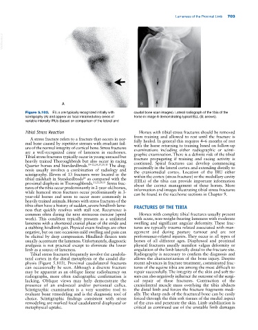
Figure 5.102. ELLs are typically recognized initially with caudal bone scan images). Lateral radiograph of the tibia of the
scintigraphy (A) and appear as focal intramedullary areas of horse in image A demonstrating typical ELL (B; arrows).
variable intensity IRUs (based on comparison of the lateral and
Tibial Stress Reaction Horses with tibial stress fractures should be removed
from training and allowed to rest until the fracture is
A stress fracture refers to a fracture that occurs in nor-
mal bone caused by repetitive stresses with resultant fail- fully healed. In general this requires 4–6 months of rest
with the horse returning to training based on follow‐up
ure of the normal integrity of cortical bone. Stress fractures examinations including either radiographic or scinti-
are a well‐recognized cause of lameness in racehorses. graphic examination. There is a definite risk of the tibial
Tibial stress fractures typically occur in young unraced but fracture propagating if training and racing activity is
heavily trained Thoroughbreds but also occur in racing continued. Spiral fractures can develop commencing
Quarter horses and Standardbreds. 19–22,24,25,28,31 The diag- proximally in the lateral cortex and extending distally to
nosis usually involves a combination of radiology and the craniomedial cortex. Location of the IRU either
scintigraphy. Eleven of 13 fractures were located in the within the cortex (stress fracture) or the medullary cavity
tibial midshaft in Standardbreds as compared with the (EELs) of the tibia can provide important information
24
proximal diaphysis in Thoroughbreds. 21,22,28,31 Stress frac- about the correct management of these horses. More
tures of the tibia occur predominantly in 2‐year‐old horses, information and images illustrating tibial stress fractures
while humeral stress fractures occur predominantly in 3‐ can be found in the racehorse sections in Chapter 9.
year‐old horses and seem to occur more commonly in
heavily trained animals. Horses with stress fractures of the
tibia often have a history of sudden, severe hindlimb lame- FRACTURES OF THE TIBIA
ness that quickly resolves with stall rest. Recurrence is
common often during the next strenuous exercise (speed Horses with complete tibial fractures usually present
work). This condition typically presents as a unilateral with acute, non‐weight‐bearing lameness with moderate
lameness with a shortened cranial phase of the stride and swelling and significant angular deformity. These frac-
a stabbing hindlimb gait. Physical exam findings are often tures are typically trauma related associated with man-
negative, but on rare occasions mild swelling and pain can agement and during pasture turnout and are not
be elicited by deep compression. Hindlimb flexion tests performance‐related injuries. They occur in all types of
usually accentuate the lameness. Unfortunately, diagnostic horses of all different ages. Diaphyseal and proximal
analgesia is not practical except to eliminate the lower physeal fractures usually manifest valgus deformity or
limb as a source of lameness. angulation of the limb laterally distal to the fracture site.
Tibial stress fractures frequently involve the caudola- Radiography is necessary to confirm the diagnosis and
teral cortex in the distal metaphysis or the caudal dia- allows the characterization of the bone injury. Despite
physis (Figure 5.103). Proximal caudolateral fractures recent advances in fracture treatment, comminuted frac-
can occasionally be seen. Although a discrete fracture tures of the equine tibia are among the most difficult to
may be apparent as an oblique linear radiolucency on repair successfully. The integrity of the skin and soft tis-
radiographs, more often radiographic confirmation is sues can also negatively influence the outcome of the surgi-
lacking. Oblique views may help demonstrate the cal repair of these fractures. Contraction of the
presence of an endosteal and/or periosteal callus. craniolateral muscle mass overlying the tibia abducts
Scintigraphic examination is a very sensitive tool to the distal limb and forces the fracture fragments medi-
evaluate bone remodeling and is the diagnostic tool of ally. The sharp ends of the fractured bone can be easily
choice. Scintigraphic findings consistent with stress forced through the thin soft tissues of the medial aspect
remodeling are marked focal caudolateral diaphyseal or of the crus and penetrate the skin. Limb stabilization is
metaphyseal uptake. critical as continued use of the unstable limb damages