
Page 738 - Adams and Stashak's Lameness in Horses, 7th Edition
P. 738
704 Chapter 5
VetBooks.ir
A B
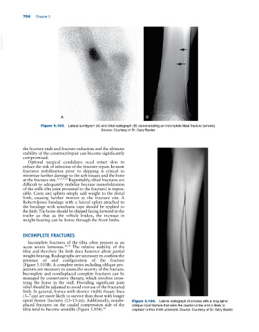
Figure 5.103. Lateral scintigram (A) and tibial radiograph (B) demonstrating an incomplete tibial fracture (arrows).
Source: Courtesy of Dr. Gary Baxter.
the fracture ends and fracture reduction and the ultimate
stability of the construct/repair can become significantly
compromised.
Optimal surgical candidates need intact skin to
reduce the risk of infection of the fracture repair. In most
fractures stabilization prior to shipping is critical to
minimize further damage to the soft tissues and the bone
at the fracture site. 3,5,7,9,32 Regrettably, tibial fractures are
difficult to adequately stabilize because immobilization
of the stifle (the joint proximal to the fracture) is impos-
sible. Casts and splints simply add weight to the distal
limb, causing further motion at the fracture site. A
Robert‐Jones bandage with a lateral splint attached to
the bandage with nonelastic tape should be applied to
the limb. The horse should be shipped facing forward in the
trailer so that as the vehicle brakes, the increase in
weight‐bearing can be borne through the front limbs.
INCOMPLETE FRACTURES
Incomplete fractures of the tibia often present as an
acute severe lameness. 15,16 The relative stability of the
tibia and therefore the limb does however allow partial
weight‐bearing. Radiographs are necessary to confirm the
presence of and configuration of the fracture
(Figure 5.103B). A complete series including oblique pro-
jections are necessary to assess the severity of the fracture.
Incomplete and nondisplaced complete fractures can be
managed by conservative therapy, which involves cross‐
tying the horse in the stall. Providing significant pain
relief should be adjusted to avoid overuse of the fractured
limb. In general, horses with shorter visible fissure lines
(3–7 cm) are more likely to survive than those with longer
spiral fissure fractures (12–15 cm). Additionally, nondis- Figure 5.104. Lateral radiograph of a horse with a long spiral
placed fractures on the caudal compression side of the oblique tibial fracture that exits the caudal cortex and is likely to
tibia tend to become unstable (Figure 5.104). 10 displace further if left untreated. Source: Courtesy of Dr. Gary Baxter.