
Page 743 - Adams and Stashak's Lameness in Horses, 7th Edition
P. 743
Lameness of the Proximal Limb 709
THE STIFLE: FEMOROPATELLAR REGION
VetBooks.ir gary m. baxter and Ken e. sullins
INTRODUCTION horses, often retain mild femoropatellar effusion with-
out a clinical problem being present. Femoropatellar
The stifle is the largest and most complex joint in the effusion can be secondary to a problem in the MFT
horse, and not surprisingly injury to the stifle is an joint, but is often less severe than with primary FP joint
important cause of hindlimb lameness. The stifle con- involvement. Femoropatellar effusion may be simple
sists of three synovial compartments (the femoropatellar fluid distension or there may be thickening of the periar-
[FP] joint, the medial femorotibial [MFT] joint, the ticular soft tissues. Such thickening may be edema or
lateral femorotibial [LFT] joint), three individual patel- consist of fibrosis from chronic inflammation. With
lar ligaments, medial and lateral collateral ligaments, chronic lameness, atrophy of the gluteal and quadriceps
and a complex array of intrasynovial ligaments and muscles on the affected side may be apparent. This may
menisci that are necessary to support the function of the be obvious, or careful comparison from the rear and
joints. The reader is referred to Chapter 1 for more side may be necessary. Hindlimb flexion, either upper
detailed anatomy of the stifle region. limb or full limb flexion, induces a painful response in
Historically, stifle problems in horses have been most cases.
reported to represent between 2% and 8% of horses Stifle pain causes typical hindlimb lameness. Viewed
presenting for lameness. 4,99 In one series of 553 horses from the side, the cranial phase of the stride is short-
with hindlimb lameness, 326 of 795 stifles that were ened, and the foot is carried closer to the ground. The
radiographed had visible abnormalities. Femoropatellar toe may drag when the horse advances the limb at a trot,
87
and femorotibial lesions occurred at similar rates of and toe wear may be obvious. When viewed from the
27% and 32%, respectively, and there was an overall rear at a trot, asymmetry in pelvic movement is often
incidence of 32% with evidence of osteoarthritis (OA). observed. The duration of gluteal rise is shorter, result-
In general, clinical problems involve the FP and MFT ing in an early unweighting of the lame limb. This often
joints more commonly than the LFT joint in athletic results in a pelvic rise on the lame limb, which can often
horses. Regardless of the type of horse, stifle problems be seen best when viewed from the side (the reader is
appear to be quite common in routine referral practice referred to Chapter 2 for more details on the lameness
and may increase as more advanced imaging capabilities exam). The degree of lameness varies according to the
continue to improve our abilities to make specific severity of the injury. Stifle lameness usually cannot be
diagnoses. 7,60,67 definitively distinguished from hock pain or other sites
of pain in the hindlimb. In some cases the horse may
Clinical Findings and Diagnostics
The evaluation of stifle lameness is made by visual
observation, palpation of the joints, gait evaluation, and
elimination of other types of lameness. The examiner
should become acquainted with normal palpation and
normal variations; asymmetry within the stifle usually
indicates a problem. Swelling may be impressive with
acute injuries, especially with extracapsular swelling,
which complicates the ability to make precise anatomic
characterization. Acutely painful horses usually do not
bear full weight by fixing the limb in extension when
walking or standing. Bruising from external trauma is
common from being kicked and in horses that jump
over fixed fences. Local and systemic anti‐inflammatory
therapy may be required to reduce the swelling before a
complete diagnosis can be made, although improve-
ments in diagnostic imaging, namely, diagnostic ultra-
sound, can make characterization possible. 7,27,28
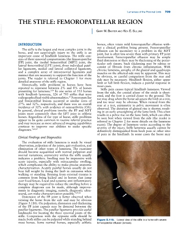
Distension of the FP joint is better observed when
viewing the horse from the side and may be obvious
(Figure 5.110). On palpation, distension and thickening
of the FP joint capsule may be detected between the
patellar ligaments. The patellar ligaments provide useful
landmarks for locating the three synovial joints of the
stifle. Comparison with the opposite stifle should be
made; both stifles can be palpated while standing behind Figure 5.110. Lateral view of the stifle in a horse with severe
most horses. Some normal horses, especially athletic femoropatellar effusion (arrows).