
Page 741 - Adams and Stashak's Lameness in Horses, 7th Edition
P. 741
Lameness of the Proximal Limb 707
diffuse and crepitus is often palpable over the tibial crest GASTROCNEMIUS DISRUPTION
(Figure 5.107). These fractures can be articular or nonar- IN FOALS AND ADULTS
VetBooks.ir proximally. Synovial effusion often accompanies frac- function of the reciprocal apparatus. Gastrocnemius mus-
ticular with the fragment frequently being displaced
Disruption of the gastrocnemius muscle can cause dys-
tures with articular components, but the effusion may be
difficult to delineate from the overall swelling. A com- cle injury is rare but can be a source of lameness in the
plete radiographic study of the stifle is necessary to accu-
rately define the fracture. The oblique orientation of the
tibial crest requires an oblique Cd35″L‐CrMO radio-
graphic view as the most useful in visualizing the fracture
line and determining the fracture configuration.
The most common fracture of the tibial tuberosity is
a nondisplaced nonarticular fracture. These fractures
can heal satisfactorily with conservative management in
the form of stall confinement and cross‐tying. Careful
radiographic monitoring is critical to assess fracture
fragment displacement and to monitor healing. Most
nondisplaced fractures with adequate stability are suc-
cessfully managed and do not require internal fixation.
2
Stall confinement for a period of 60 days with serial
radiographs to confirm appropriate healing is used to
determine when exercise can begin. Smaller fracture
fragments that are associated with wounds may require
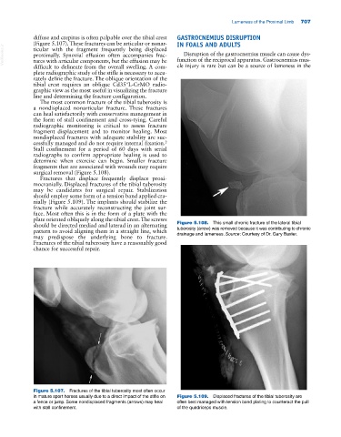
surgical removal (Figure 5.108).
Fractures that displace frequently displace proxi-
mocranially. Displaced fractures of the tibial tuberosity
may be candidates for surgical repair. Stabilization
should employ some form of a tension band applied cra-
nially (Figure 5.109). The implants should stabilize the
fracture while accurately reconstructing the joint sur-
face. Most often this is in the form of a plate with the
plate oriented obliquely along the tibial crest. The screws
should be directed mediad and laterad in an alternating Figure 5.108. This small chronic fracture of the lateral tibial
pattern to avoid aligning them in a straight line, which tuberosity (arrow) was removed because it was contributing to chronic
may predispose the underlying bone to fracture. drainage and lameness. Source: Courtesy of Dr. Gary Baxter.
Fractures of the tibial tuberosity have a reasonably good
chance for successful repair.
Figure 5.107. Fractures of the tibial tuberosity most often occur
in mature sport horses usually due to a direct impact of the stifle on Figure 5.109. Displaced fractures of the tibial tuberosity are
a fence or jump. Some nondisplaced fragments (arrows) may heal often best managed with tension band plating to counteract the pull
with stall confinement. of the quadriceps muscle.