
Page 736 - Adams and Stashak's Lameness in Horses, 7th Edition
P. 736
702 Chapter 5
puncture wounds. More extensive injuries may involve become open. Younger animals are subjected to the same
the tibia. Fractures can occur in about the same fre- types of traumatic injuries as adults, but because they are
VetBooks.ir incomplete, proximal physeal fractures (in young involve the growth plate(s). As in all long bone fractures
actively growing, fracture configurations will often
quency as other long bones. Fracture types include
in horses, maintaining the integrity of the skin and appro-
horses), diaphyseal fractures, and tibial crest frac-
tures. 2–13,15–18,26,27,29,34–36 Training‐related bone injuries priately stabilizing the limb before shipping are critical to
also occur in the shaft of the tibia. 1,19–22,24,25,28,31 In race- a successful outcome if surgery is being considered. 5
horses stress‐related bone injury occurs in the tibial
cortex and often present as a subtle, high‐speed lame-
ness. In addition, enostosis‐like lesions (ELLs), similar to ENOSTOSIS‐LIKE LESIONS
what is seen in humans, have been described in horses
and occur as focal or multifocal sclerotic lesions within ELLs were first described in horses as focal or multi-
the medullary cavity of long bones. 1 focal sclerotic lesions within the medullary cavity of the
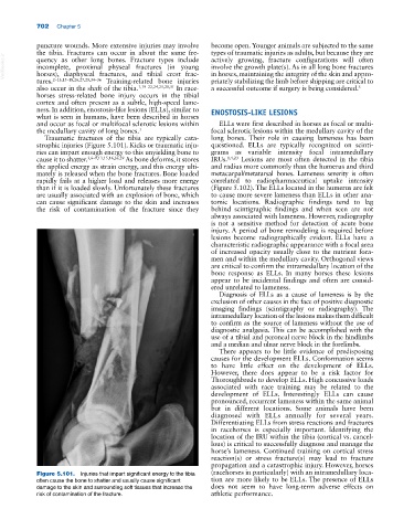
Traumatic fractures of the tibia are typically cata- long bones. Their role in causing lameness has been
strophic injuries (Figure 5.101). Kicks or traumatic inju- questioned. ELLs are typically recognized on scinti-
ries can impart enough energy to this unyielding bone to grams as variable intensity focal intramedullary
cause it to shatter. 3,6–9,13,15,16,26,29 As bone deforms, it stores IRUs. 1,4,23 Lesions are most often detected in the tibia
the applied energy as strain energy, and this energy ulti- and radius more commonly than the humerus and third
mately is released when the bone fractures. Bone loaded metacarpal/metatarsal bones. Lameness severity is often
rapidly fails at a higher load and releases more energy correlated to radiopharmaceutical uptake intensity
than if it is loaded slowly. Unfortunately these fractures (Figure 5.102). The ELLs located in the humerus are felt
are usually associated with an explosion of bone, which to cause more severe lameness than ELLs in other ana-
can cause significant damage to the skin and increases tomic locations. Radiographic findings tend to lag
the risk of contamination of the fracture since they behind scintigraphic findings and when seen are not
always associated with lameness. However, radiography
is not a sensitive method for detection of acute bone
injury. A period of bone remodeling is required before
lesions become radiographically evident. ELLs have a
characteristic radiographic appearance with a focal area
of increased opacity usually close to the nutrient fora-
men and within the medullary cavity. Orthogonal views
are critical to confirm the intramedullary location of the
bone response as ELLs. In many horses these lesions
appear to be incidental findings and often are consid-
ered unrelated to lameness.
Diagnosis of ELLs as a cause of lameness is by the
exclusion of other causes in the face of positive diagnostic
imaging findings (scintigraphy or radiography). The
intramedullary location of the lesions makes them difficult
to confirm as the source of lameness without the use of
diagnostic analgesia. This can be accomplished with the
use of a tibial and peroneal nerve block in the hindlimbs
and a median and ulnar nerve block in the forelimbs.
There appears to be little evidence of predisposing
causes for the development ELLs. Conformation seems
to have little effect on the development of ELLs.
However, there does appear to be a risk factor for
Thoroughbreds to develop ELLs. High concussive loads
associated with race training may be related to the
development of ELLs. Interestingly ELLs can cause
pronounced, recurrent lameness within the same animal
but in different locations. Some animals have been
diagnosed with ELLs annually for several years.
Differentiating ELLs from stress reactions and fractures
in racehorses is especially important. Identifying the
location of the IRU within the tibia (cortical vs. cancel-
lous) is critical to successfully diagnose and manage the
horse’s lameness. Continued training on cortical stress
reaction(s) or stress fracture(s) may lead to fracture
propagation and a catastrophic injury. However, horses
Figure 5.101. Injuries that impart significant energy to the tibia (racehorses in particularly) with an intramedullary loca-
often cause the bone to shatter and usually cause significant tion are more likely to be ELLs. The presence of ELLs
damage to the skin and surrounding soft tissues that increase the does not seem to have long‐term adverse effects on
risk of contamination of the fracture. athletic performance.