
Page 800 - Adams and Stashak's Lameness in Horses, 7th Edition
P. 800
766 Chapter 6
Clinical Examination head allows assessment of the amount of dorsiflexion as
well as the symmetry of the muscles and location of ana-
Clinical examination of the horse is performed as
VetBooks.ir with any lameness case, with more emphasis on obser- (Figure 6.2).
tomical landmarks on the ventral aspect of the neck
vation of lateral bending in walking serpentines and cir-
In the thorax, a similar manipulation (pulling on the
cles at the walk, trot, and canter. The quality of the walk
(4‐beat) and canter (3‐beat) can give essential informa- tail with one hand and placing the other arm as a ful-
crum at an individual horizontal process of a specific
tion about location of a lesion within the spine due to vertebra) provides information about the range of
the specific loads that are generated by these gaits to motion and intervertebral mobility in the lateral plane
specific parts of the spine (Table 6.1). (Figure 6.3). Palpation of the withers, dorsal processes
In the walk and trot in hand on a straight line, obser- of the thoracic vertebrae, and associated muscles indicate
vation of the gait and the position of the hindlimbs in pain in this region that is very susceptible to discomfort
relation to the front limbs and the position of the tail caused by poor saddle fit. Triggering the dorsiflexion
can assist in assessing for the presence and location of reflex by pressure with a pen in a paramedian motion
back pain. For example, do the hindlimbs track the fore-
limbs, and are the haunches more to one side? Because
there is more mobility of the lumbosacral spine in the
canter, lumbosacral pain is more obvious at this gait. By
contrast, thoracic pain appears more as stiffness in the
trot and resistance to going downhill, or in making
downward transitions from canter to trot or trot to
walk. More detailed signs and the locations they may
identify are presented in Table 6.2.
Information gleaned from palpation of the back can
be confusing. Most horses with any kind of back pain
exhibit some muscle tension in the epaxial muscles. It is
nearly impossible to differentiate between muscle ten-
sion caused by guarding spinal structures and primary
muscle tension due to muscle problems. See the section
on muscle diseases in Chapter 7.
Sensitivity to touch or pressure is another symptom
that can be difficult to interpret. Some horses initially
show a defensive reaction to every contact with their
back, even when there is no pathological condition pre-
sent. To make an initial differentiation, touch or apply
pressure very gently and when the horse resists, main-
tain it and wait for the initial reaction to fade. In behav-
ioral issues, when the pressure persists without correction
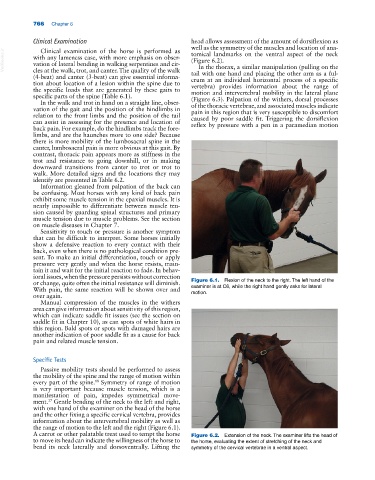
or change, quite often the initial resistance will diminish. Figure 6.1. Flexion of the neck to the right. The left hand of the
With pain, the same reaction will be shown over and examiner is at C6, while the right hand gently asks for lateral
over again. motion.
Manual compression of the muscles in the withers
area can give information about sensitivity of this region,
which can indicate saddle fit issues (see the section on
saddle fit in Chapter 10), as can spots of white hairs in
this region. Bald spots or spots with damaged hairs are
another indication of poor saddle fit as a cause for back
pain and related muscle tension.
Specific Tests
Passive mobility tests should be performed to assess
the mobility of the spine and the range of motion within
every part of the spine. Symmetry of range of motion
18
is very important because muscle tension, which is a
manifestation of pain, impedes symmetrical move-
ment. Gentle bending of the neck to the left and right,
17
with one hand of the examiner on the head of the horse
and the other fixing a specific cervical vertebra, provides
information about the intervertebral mobility as well as
the range of motion to the left and the right (Figure 6.1).
A carrot or other palatable treat used to tempt the horse Figure 6.2. Extension of the neck. The examiner lifts the head of
to move its head can indicate the willingness of the horse to the horse, evaluating the extent of stretching of the neck and
bend its neck laterally and dorsoventrally. Lifting the symmetry of the cervical vertebrae in a ventral aspect.