
Page 801 - Adams and Stashak's Lameness in Horses, 7th Edition
P. 801
Lameness Associated with the Axial Skeleton 767
VetBooks.ir
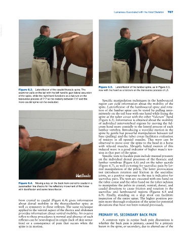
Figure 6.5. Lateroflexion of the lumbar spine, as in Figure 6.3,
Figure 6.3. Lateroflexion of the caudal thoracic spine. The now with the hand as a fulcrum on the transverse process of L3.
examiner pulls on the tail with the left hand to gain lateral excursion
of the spine, while the right hand functions as a fulcrum on the
transverse process of T17 so the mobility between T17 and the Specific manipulation techniques in the lumbosacral
more caudal spine can be evaluated.
region can yield information about the mobility of the
spine. Lateroflexion of the lumbosacral spine and rota-
tion of the lumbar spine can be tested by pulling inter-
mittently on the tail base with one hand while fixing the
spine at the tuber coxae with the other “fulcrum” hand
(Figure 6.5). Information is obtained about the mobility
of individual intervertebral regions by moving the ful-
crum hand more cranially to the lateral process of each
lumbar vertebra. Introducing a wavelike motion in the
spine by gentle but powerful manipulation between tail
base (pulling) and the tuber coxae facilitates evaluation
of tension in all epaxial muscles. This wave can be
observed to move over the spine to the head in a horse
with relaxed muscles. Abruptly halted motion of this
induced wave is a good indicator of higher muscle ten-
sion in that part of the spine.
Specific tests to localize pain include manual pressure
on the individual dorsal processes of the thoracic and
lumbar vertebrae (Figure 6.6) and on the tuber sacrale
(Figure 6.7), as well as testing for sacroiliac pain by lat-
eral manipulation of the pelvis. The latter provocation
test introduces rotation and friction in the sacroiliac
joints, so a positive response to the test is indicative for
sacroiliac pain. The tests are executed with one hand on
the tuber coxae and the other hand on the tuber ischium
Figure 6.4. Moving a key on the back from cranial to caudal in a
paramedian line checks for the reflectory movement of the horse to manipulate the pelvis in cranial, ventral, dorsal, and
with dorsiflexion and some lateroflexion. caudal directions to cause friction and rotation in the
sacroiliac and lumbosacral regions (Figures 6.8 and
6.9). Finally, standing on a step stool can facilitate
inspection of the entire spine. The higher position per-
from cranial to caudal (Figure 6.4) gives information mits more thorough evaluation of the spine for potential
about dorsal mobility in the thoracolumbar spine as deviations that have not been noticed previously.
well as symmetry in these reflexes. The same technique
applied to the ventral aspect of the thorax and abdomen
provides information about ventral mobility. An evasive PRIMARY VS. SECONDARY BACK PAIN
reflex to these procedures is normal and absence of such
reflexes can be neurological in origin (lack of skin sensa- A common topic in equine back pain discussions is
tion) or a consequence of pain that occurs when the whether the back pain is primary, caused by a primary
spine is in motion. lesion in the spine, or secondary, due to altered use of the