
Page 806 - Adams and Stashak's Lameness in Horses, 7th Edition
P. 806
772 Chapter 6
VetBooks.ir
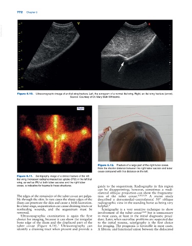
Figure 6.10. Ultrasonographic image of an ilial wing fracture. Left, the sonogram of a normal ilial wing. Right, an ilial wing fracture (arrow).
Source: Courtesy of Dr. Mary Beth Whitcomb.
Figure 6.12. Fracture of a large part of the right tuber coxae.
Note the shorter distance between the right tuber sacrale and tuber
coxae compared with this distance on the left.
Figure 6.11. Scintigraphy image of a stress fracture of the left
ilial wing. Increased radiopharmaceutical uptake (IRU) in the left ilial
wing, as well as IRU at both tuber sacrales and the right tuber
coxae, is indicative for trauma to these structures. guide to the sequestrum. Radiography in this region
can be disappointing; however, sometimes a medi-
olateral oblique projection can show the fragmenta-
The edges of the remainder of the tuber coxae are palpa- tion of the tuber coxae. 8,18,20,26 A recent study
ble through the skin. In rare cases the sharp edges of the described a dorsomedial–centrolateral 50° oblique
ilium can penetrate the skin and cause a fresh laceration. radiographic view in the standing horse as being very
In a later stage, sequestration can cause draining tracts or helpful. 8
nonhealing wounds, and the sequestrum must be Scintigraphy is a very sensitive technique to show
removed. involvement of the tuber coxae 12,20 but is unnecessary
Ultrasonographic examination is again the first in most cases, at least in the initial diagnostic proce-
choice for imaging, because it can show the irregular dure. Later, when sacroiliac problems are suspected due
bone edge of the ilium and the displaced part of the to the initial trauma, scintigraphy is the first choice
tuber coxae (Figure 6.14). Ultrasonography can for imaging. The prognosis is favorable in most cases.
1
identify a draining tract when present and provide a A fibrotic and functional union between the dislocated