
Page 807 - Adams and Stashak's Lameness in Horses, 7th Edition
P. 807
Lameness Associated with the Axial Skeleton 773
VetBooks.ir
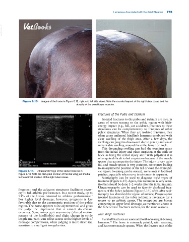
Figure 6.13. Images of the horse in Figure 6.12, right and left side views. Note the rounded aspect of the right tuber coxae and the
atrophy of the quadriceps muscles.
Fractures of the Pubis and Ischium
Isolated fractures to the pubis and ischium are rare. In
cases of severe trauma to the pelvic region with high‐
energy impact (e.g., fall, car accident), fractures to these
structures can be complementary to fractures of other
pelvic structures. When they are isolated fractures, they
often cause unilateral hindlimb lameness combined with
clear swelling of the thigh area. After a few days, the
swelling can progress downward due to gravity and cause
remarkable swelling around the stifle, femur, or hock.
This descending swelling can lead the examiner away
from the initial injury and place suspicion at the stifle or
hock as being the initial injury site. With palpation it is
5
often quite difficult to feel crepitation because of the muscle
spasm that accompanies the injury. The injury is very pain-
ful, and muscle spasm is very common, sometimes leading
to an asymmetric position of the tail or even the entire pel-
Figure 6.14. Ultrasound image of the same horse as in vic region. Sweating can be noticed, sometimes in localized
Figure 6.13. Note the disrupted contour of the ilial wing just medial patches, especially when nerve involvement is apparent.
to the normal position of the right tuber coxae. Scintigraphy can be used to diagnose these types of
fractures (Figure 6.15). Again, this technique is very sensi-
tive but should be done 1–2 weeks after the initial trauma.
Ultrasonography can be used to identify displaced frag-
fragment and the adjacent structures facilitates recov- ments of the tuber ischium (Figure 6.16), often after scin-
ery to full athletic performance. In a recent study, up to tigraphy has identified the region of interest. Prognosis for
93% of the horses returned to athletic performance. isolated fractures of the tuber ischium is favorable for a
8
For higher level dressage, however, prognosis is less return to an athletic career. The exceptions are horses
favorable due to the asymmetric position of the pelvic competing in upper level dressage, as mentioned above in
region. The horse appears to be asymmetrical and gives the tuber coxae fractures section, for the same reasons.
the judge the impression that it cannot do certain
exercises. Some minor gait asymmetry (altered motion
pattern of the hindlimb(s) and slight change in stride Ilial Shaft Fractures
length and path) can affect scores at the higher levels of Ilial shaft fractures are associated with non‐weight‐ bearing
dressage competitions, when judging is more strict and lameness. The horse is extremely painful, with sweating,
24
sensitive to small gait irregularities. and has severe muscle spasms. When the fracture ends of the