
Page 812 - Adams and Stashak's Lameness in Horses, 7th Edition
P. 812
778 Chapter 6
and semitendinosus muscles, and at the ventral part of
the pelvis are the psoas muscles, all of which are impor-
VetBooks.ir Dorsal sacral ligament the muscles in this region maintain the mobility of the
tant contributors to stability. Under normal conditions,
sacroiliac joints within their physical limits. However,
when the muscles are not powerful enough to compete
with the external forces that result from massive trauma
due to falling, slipping, tipping over, etc., primary liga-
ment damage and primary or secondary joint trauma
can be sustained. Weakness in these muscles can be asso-
ciated with sacroiliac injury. This weakness can have
many causes, including fatigue from improper training
techniques, overuse, and repetitive stress injuries that
can occur in young horses not yet capable of the desired
level of performance. 18
The pelvic region is not a rigid structure, as was previ-
16
Tuber sacrale ously thought. Haussler et al. discovered that there is a
small amount of deformation of the pelvic bone struc-
tures during the loading cycle in motion, and all of the
involved structures (the bone, ligamentous structures, and
muscles) form a semiflexible complex to assist propulsion
and transport this propulsion from the hindlimbs onto
the spinal column. These findings make it easier to
10
understand the major impact of the dysfunction of the
pelvis structures regarding locomotion and performance.
Three different types of injury occur in the sacroiliac
region. In the author’s experience, the most common
seem to be those that cause damage to the dorsal sacral
ligaments (the structure with the greatest span between
tuber sacrale, sacral bone, and the sacrosciatic ligament
and the most potential motion between these structures).
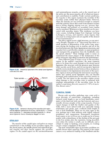
Figure 6.22. Anatomical specimen of the dorsal sacral ligament,
cross‐sectional view. When trauma to the ligamentous structures of the sacro-
iliac region is more profound, the interosseus sacral liga-
ments and ventral sacral ligaments also can become
damaged, with (sub)luxation of the sacroiliac joint(s) as
3
Tuber sacrale Sacrum a result. The last injury to the sacroiliac region occurs
when damage to the sacroiliac joint is sustained with
resultant osteoarthritis of the sacroiliac joint. In the
author’s experience, this only occurs in 5–10% of sacro-
2 2 iliac disease cases. 24
Sacral wing
1 1 CLINICAL SIGNS
Horses with sacroiliac pathology may come with a
great diversity of owner/rider complaints, including
reduced stride length in one or both hindlimbs, asym-
metry of the hind end with one hip lowered, and occa-
sionally obvious atrophy of the croup muscles. Owners
report changes in both the rhythm and quality of the
walk, with a more lateral walk seen especially during
Figure 6.23. Schematic drawing of the sacroiliac joint region serpentines and circles, and a reduced stride length in
and the adjacent ligamentous structures, cranial aspect. 1 = ventral one or both hindlimbs. There are no clear signs at the
sacral ligaments, 2 = interosseus sacroiliac ligaments, 3 = dorsal trot, perhaps just a little stiffness with slightly reduced
sacral ligaments. Source: Drawing by Maggie Hofmann. propulsion and engagement. Downward transitions
from canter to trot or from trot to walk can be of lower
quality with a disturbance of the rhythm of the new gait
ETIOLOGY in the first strides after the transition. Going downhill,
even when the incline is small, can be difficult, and a
The muscles of the caudal spine and pelvis are major refusal to jump in hunting, cross‐country, or eventing
contributors to the stability of the sacroiliac region. On can be a major complaint of the rider. 2,3
the dorsal aspect, the middle gluteal and superficial glu- The most remarkable signs of sacroiliac problems are
teal muscles and their fasciae support the sacroiliac usually shown when cantering. The pattern of the canter
region. On the caudal aspect are the semimembranosus causes a very unilateral loading of the hindlimb and the