
Page 814 - Adams and Stashak's Lameness in Horses, 7th Edition
P. 814
780 Chapter 6
VetBooks.ir
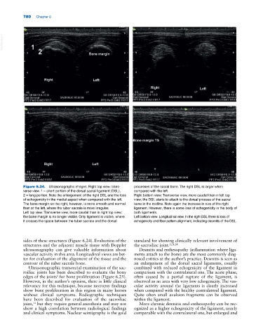
Figure 6.24. Ultrasonographic images. Right top view: trans- processes of the sacral bone. The right DSL is larger when
verse view. 1 = short portion of the dorsal sacral ligament (DSL), compared with the left.
2 = long portion. Note the enlargement of the right DSL and the loss Right bottom view: Transverse view, more caudal than in left top
of echogenicity in the medial aspect when compared with the left. view; the DSL starts to attach to the dorsal process of the sacral
The bone margin on the right, however, is more smooth and normal bone in the midline. Note again the increase in size of the right
than at the left, where the tuber sacrale is more irregular. ligament. However, there is some loss of echogenicity in the body of
Left top view: Transverse view, more caudal than in right top view; both ligaments.
the bone margin is no longer visible. Only ligament is visible, where Left bottom view: Longitudinal view. In the right DSL there is loss of
it crosses the space between the tuber sacrale and the dorsal echogenicity and fiber pattern alignment, indicating desmitis of the DSL.
sides of these structures (Figure 6.24). Evaluation of the standard for showing clinically relevant involvement of
structures and the adjacent muscle tissue with Doppler the sacroiliac joint. 3,21,24
ultrasonography can give valuable information about Desmitis and enthesopathy (inflammation where liga-
vascular activity in this area. Longitudinal views are bet- ments attach to the bone) are the most commonly diag-
ter for evaluation of the alignment of the tissue and the nosed entities at the author’s practice. Desmitis is seen as
contour of the tuber sacrale bone. an enlargement of the dorsal sacral ligaments, usually
Ultrasonographic transrectal examination of the sac- combined with reduced echogenicity of the ligament in
roiliac joints has been described to evaluate the bony comparison with the contralateral one. The acute phase,
edges of the joints for bone proliferation (Figure 6.25). often caused by a partial rupture of the ligament, is
6
However, in the author’s opinion, there is little clinical observed as an area with very low echogenicity. The vas-
relevancy for this technique, because necropsy findings cular activity around the ligaments is clearly increased
show bone proliferation in this region in many horses when compared with the healthy contralateral ligament.
without clinical symptoms. Radiographic techniques Quite often small avulsion fragments can be observed
have been described for evaluation of the sacroiliac within the ligament.
joint, but they require general anesthesia and may not More chronic desmitis and enthesopathy can be rec-
11
show a high correlation between radiological findings ognized as a higher echogenicity of the ligament, nearly
and clinical symptoms. Nuclear scintigraphy is the gold comparable with the contralateral one, but enlarged and