
Page 815 - Adams and Stashak's Lameness in Horses, 7th Edition
P. 815
Lameness Associated with the Axial Skeleton 781
with an irregular and less clearly defined bone margin. In the case of significant displacement of the sacral
This is more a repetitive stress injury than an acute bone, the incongruence in the articular surfaces of the
VetBooks.ir structures, a more complete displacement of the sacral also can be a consequence of sacroiliac instability due to
sacroiliac joints leads to osteoarthritis. Osteoarthritis
trauma.
In cases with massive damage to the ligamentous
chronic desmitis of the dorsal sacral ligaments.
bone is visible with the ilial wings displaced dorsally and Scintigraphy facilitates evaluation of the sacroiliac
the sacral bone displaced ventrally. In this case, one or joints as well as identification of enthesopathy of the
both of the tuber sacrales are more pronounced tuber sacrale and the dorsal processes of the sacral
(Figure 6.26). The latter condition is often called “hunt- bone. 3,21 Lateral (Figure 6.27), dorsoventral, and oblique
er’s bump.” views of the sacroiliac region pinpoint increased bone
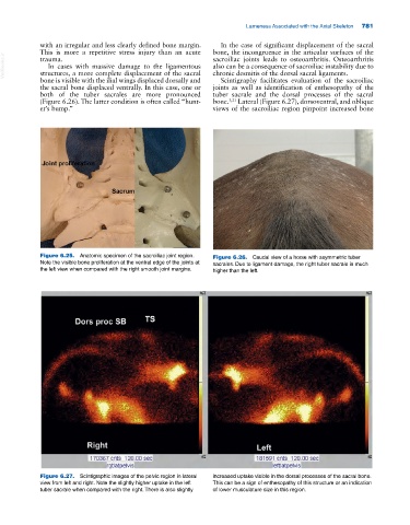
Figure 6.25. Anatomic specimen of the sacroiliac joint region. Figure 6.26. Caudal view of a horse with asymmetric tuber
Note the visible bone proliferation at the ventral edge of the joints at sacrales. Due to ligament damage, the right tuber sacrale is much
the left view when compared with the right smooth joint margins. higher than the left.
Figure 6.27. Scintigraphic images of the pelvic region in lateral increased uptake visible in the dorsal processes of the sacral bone.
view from left and right. Note the slightly higher uptake in the left This can be a sign of enthesopathy of this structure or an indication
tuber sacrale when compared with the right. There is also slightly of lower musculature size in this region.