
Page 820 - Adams and Stashak's Lameness in Horses, 7th Edition
P. 820
786 Chapter 6
Clinical Signs Treatment
Signs of desmopathy of the supraspinous ligament
As with any ligamentous structure, treatment is focused
VetBooks.ir are similar to those of impingement of dorsal spinous on rest, medication, and rehabilitation. Controlled stretch-
ing of the ligament with a lower position of the head (i.e.,
processes. When only ligament pathology is present,
signs can be mild and very difficult to detect.
lower head and a longer neck) and a gradual increase in
Occasionally owners may recognize the increased size in pasture, with hay on the ground, when ridden with a
of the ligament (focal thickening), and this can be the the workload is recommended.
primary complaint. In cases with more swelling in and
around the ligament, the appearance of the shape of
the spine can resemble kyphosis. In that case, radiog- Prognosis
raphy shows a normal configuration of the spine, but When the ligament is the only affected structure and
ultrasonography can show the soft tissue involvement adequate time for a complete recovery before resuming
and edema. the original work is allowed (4–6 months), the progno-
sis is usually favorable. When more structures are
Diagnosis involved, as with impingement of dorsal spinous pro-
cesses, facet joints, or intervertebral discs, the prognosis
Ultrasonography is the tool of choice to image these tends to be less favorable.
injuries. Diagnosis of the pathology of the supraspinous
22
ligament can be made with ultrasonographic examina-
tion. Radiography and scintigraphy can give additional
information about the involvement of the spinous FRACTURES OF THE SPINOUS PROCESSES
processes. Radiographic examination of desmopathy of
the supraspinous ligament may show irregular bone Etiology
margins of the summit of the dorsal spinal processes, Fractures of the dorsal spinous processes occur
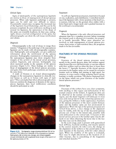
avulsion fragments, and sclerosis. Scintigraphy may mostly in the cranial thoracic spine (the withers region)
show increased uptake in the summits of the spinous when horses flip over, fall backwards, or run into objects
processes (Figure 6.31). These findings are similar to with their withers (such as when the door is lower than
those of impingement of the dorsal spinous processes, the horse). 3,24 Sporadic fractures of the dorsal spinous
and quite often both pathologic conditions are found in processes in the lumbar spine can be caused by excessive
the same horse. trauma such as falling and turning at high speed, for
A study of Henson et al. found ultrasonographic instance, in cross‐country riding, jumping, barrel racing,
changes in the supraspinous ligament in clinically nor- hunting, or traffic accidents. This places rotational force
mal horses as well as in horses with signs of back on the spine, which can cause fractures of the lumbar
pain. They concluded that when ultrasonography alone lateral spinous processes. 19
22
does not confirm the diagnosis of desmitis of the supras-
pinous ligament, then local (infiltration) anesthesia
should be performed as well. Clinical Signs
Fractures of the withers have very clear symptoms,
with swelling in this region and deformation of the
normal contour of the withers. A significant dent can
be observed in the normal curvature of the withers
region, and the distal fragment will dislocate in most
cases and end up lateral to the remaining spinous pro-
cess, giving the withers a wider appearance. Fractures
of the spinous processes of the more caudal thoracic
spine and the lumbar spine are less pronounced in
appearance. When the tips of the spinous processes are
viewed from above, a clear change in the alignment
may be visible, with the tip of the fractured spinous
process out of line to the lateral side, possibly with
some dislocation to the ventral side. A fracture of the
lateral spinous process in the lumbar spine may be dif-
ficult to see when observing the horse due to the fact
that they may not be dislocated.
In the acute stage, pain is present with generalized
back stiffness, localized swelling, and muscle spasms
in the epaxial muscles. In older cases, the change in
the alignment of the withers or the caudal spine is the
Figure 6.31. Scintigraphic image showing mild focal IRU at the clear remains of the fracture, with few other clinical
summit of T18, indicative of desmitis of the supraspinous ligament. signs. When the mobility of the spine is evaluated,
Radiography did not show any change, and ultrasonography there may be slight alterations at the location of an
showed mild desmitis with roughened bone margin of the summit of older fracture, due to remodeling of the fragments
the spinous process of T18. and attached muscles. 20,28