
Page 821 - Adams and Stashak's Lameness in Horses, 7th Edition
P. 821
Lameness Associated with the Axial Skeleton 787
Diagnosis region, it is difficult to make the saddle fit properly due
to the changed shape of the withers. When the horse is
Radiographic examination is the first diagnostic tech-
VetBooks.ir nique to image the spinous processes because the frag- hand showing, breeding championships), the abnormal
judged on shape and conformation in its discipline (i.e.,
ments, which are often dislocated, show clearly on the
shape of the withers can influence the judging. Providing
radiographs. The summits of the thoracic spinous pro-
37
cesses, especially in the withers region (T3 to T12), can the client with a clear veterinary statement that the
abnormality is caused by a traumatic event and not
vary in shape, show a variety in the pattern of accessory genetic and that the horse is capable of competing facili-
centers of ossification, and easily be misinterpreted as a tates a successful show career.
fracture. Dislocation of the fragment is a very common
finding in fractures of the spinous processes in the with-
ers region, which helps to distinguish them from irregu- VERTEBRAL FRACTURES
larities and delayed ossification. In the more caudal
thoracic spine and lumbar spine, radiographic examina- Etiology
tion may be less useful because the muscle mass in these Most vertebral fractures are traumatic in origin;
regions can make radiographic imaging incapable of however, pathological fractures can occur with pro-
identifying a nondisplaced fracture. cesses affecting the vertebrae such as neoplasia, metasta-
Ultrasonographic examination can give good additional sis, and osteomyelitis. 34
information, especially to identify fractures of the lateral Stress fractures of the vertebra most likely occur more
spinous processes in the lumbar spine. 9 frequently than previously believed. In one study, 18 of
Scintigraphy can show increased radiopharmaceuti- 36 specimens had incomplete fractures and focal peri-
cal uptake after a fracture, but is not specific enough to osteal proliferation of the lamina stemming from verte-
identify the fractures. For lateral spinous process frac- bral stress fractures. The fractures occurred most often
19
tures, scintigraphy can be useful to identify the location near the junction of the cranial articular process and the
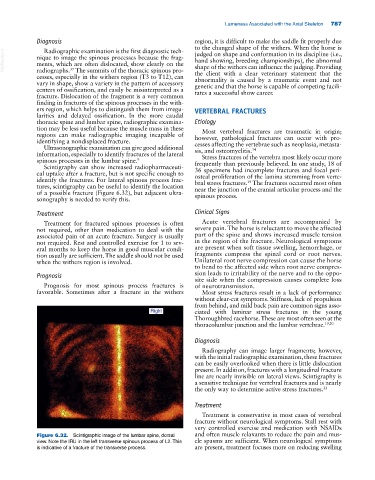
of a possible fracture (Figure 6.32), but adjacent ultra- spinous process.
sonography is needed to verify this.
Treatment Clinical Signs
Treatment for fractured spinous processes is often Acute vertebral fractures are accompanied by
not required, other than medication to deal with the severe pain. The horse is reluctant to move the affected
associated pain of an acute fracture. Surgery is usually part of the spine and shows increased muscle tension
not required. Rest and controlled exercise for 1 to sev- in the region of the fracture. Neurological symptoms
eral months to keep the horse in good muscular condi- are present when soft tissue swelling, hemorrhage, or
tion usually are sufficient. The saddle should not be used fragments compress the spinal cord or root nerves.
when the withers region is involved. Unilateral root nerve compression can cause the horse
to bend to the affected side when root nerve compres-
Prognosis sion leads to irritability of the nerve and to the oppo-
site side when the compression causes complete loss
Prognosis for most spinous process fractures is of neurotransmission.
favorable. Sometimes after a fracture in the withers Most stress fractures result in a lack of performance
without clear‐cut symptoms. Stiffness, lack of propulsion
from behind, and mild back pain are common signs asso-
ciated with laminar stress fractures in the young
Thoroughbred racehorse. These are most often seen at the
thoracolumbar junction and the lumbar vertebrae. 19,20
Diagnosis
Radiography can image larger fragments; however,
with the initial radiographic examination, these fractures
can be easily overlooked when there is little dislocation
present. In addition, fractures with a longitudinal fracture
line are nearly invisible on lateral views. Scintigraphy is
a sensitive technique for vertebral fractures and is nearly
the only way to determine active stress fractures. 33
Treatment
Treatment is conservative in most cases of vertebral
fracture without neurological symptoms. Stall rest with
very controlled exercise and medication with NSAIDs
Figure 6.32. Scintigraphic image of the lumbar spine, dorsal and often muscle relaxants to reduce the pain and mus-
view. Note the IRU in the left transverse spinous process of L2. This cle spasms are sufficient. When neurological symptoms
is indicative of a fracture of the transverse process. are present, treatment focuses more on reducing swelling