
Page 825 - Adams and Stashak's Lameness in Horses, 7th Edition
P. 825
Lameness Associated with the Axial Skeleton 791
VetBooks.ir
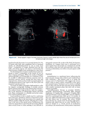
Figure 6.37. Ultrasonographic image of the lumbar facet joints. Doppler is used to identify higher blood flow around the facet joint at the
left facet joint (right in the image).
just dorsal in the facet joint. A recent study showed 61.5% adequately interpret the results of the block. In humans,
of horses with back pain complaints had an increased anesthesia of a suspect facet joint is performed on a
uptake of radiopharmaceutical in one or more facet more regular basis, but people can communicate and tell
joints. Comparison of images obtained from the left, the examiner when the main joint pain they were expe-
16
right, and dorsal aspects can assist in determining the riencing is diminished, even when a muscle spasm is still
exact location of higher bone metabolism in a facet joint. present. 31
The final diagnosis of equine vertebral facet joint syn-
drome is often a summation of the results of two or Treatment
three imaging techniques. When the results of Doppler
ultrasonography and scintigraphy are combined, it may Rehabilitation is a significant factor influencing the
be possible to differentiate between synovitis with its final outcome of facet joint arthritis and vertebral facet
increased vascular activity around the facet joint and joint syndrome. The primary goal is to break the
29
osteoarthritis, which can have both increased uptake of vicious cycle of inflammation that triggers the nerves,
radiopharmaceutical in the region of the facet joint and which leads to muscle spasm, immobility of the spine,
increased vascular activity. and repeated injury. Treatment can be aimed at multiple
The study by Gillen concluded a high predictive value spots in this cycle, but the best outcome can be expected
for negative scintigraphy findings to exclude osteoar- with complex treatment plans that deal with as many
thritis of facet joints as the cause of back pain in horses. aspects as possible.
16
The authors found a high predictive value for scintigra- Anti‐inflammatory therapy is essential. NSAIDs,
phy in the detection of radiographic lesions and back especially phenylbutazone (1 g/500 kg BID), flunixin
pain. Thus, scintigraphy is a valuable tool in the evalua- meglumine (0.5 mg/kg SID), naproxen (10 mg/kg SID),
tion of thoracolumbar pain. 16 and meclofenamic acid (2 mg/kg SID), must be adminis-
Local anesthesia is not a reliable tool for confirming tered for a prolonged time, often up to 1–2 months. In
facet joint pain. Anesthetic solution is deposited in mus- that case, additional medication to prevent gastrointesti-
cle tissue rather than in the facet joint or its capsule and nal side effects may be needed. The newer NSAID
diffuses rapidly within the muscle, affecting nerve sensa- firocoxib may be a good choice for horses with gastro-
tion in the rami of the spinal nerves. Furthermore, the intestinal side effects that are on other NSAIDs, but it
reflectory spasm that is caused by the facet joint arthritis must be limited to a 3‐week period of medication,
takes several hours to relax, making it very difficult to according to the manufacturer. Some positive experience