
Page 827 - Adams and Stashak's Lameness in Horses, 7th Edition
P. 827
Lameness Associated with the Axial Skeleton 793
symptoms can be observed. Blood analysis may reveal
10
a leukocytosis and elevated fibrinogen.
VetBooks.ir Diagnosis
Diagnosis of discospondylitis is based upon appropriate
clinical signs and radiographical findings. Radiographic
1
features of discospondylitis include lysis and/or prolifera-
tion of the vertebral bodies adjacent to the affected disc. As
the disease progresses, the intervertebral disc space may
narrow and collapse, and a smooth bony bridge may unite
36
23
the affected vertebrae. Scintigraphy, computed tomog-
raphy, and ultrasonography have been used to facilitate
35
34
the diagnosis of equine discospondylitis and vertebral
osteomyelitis. Scintigraphy may be useful to determine
whether multiple sites of bone involvement are present. 37
Treatment and Prognosis
Figure 6.39. Scintigraphic image of a horse with IRU in the
Successful treatment of discospondylitis usually midthoracic spine, suggestive of spondylosis.
involves long‐term antimicrobial administration. 2,23,36 In
specific cases surgical curettage of the lesion is an option
when access to the lesion is possible. If the causative Although the prognosis for horses with discospondy-
35
organism is not isolated, as is usually the case, broad‐ litis has generally been considered guarded, favorable
spectrum antimicrobials should be administered. outcomes have been associated with early detection,
Intravenous antimicrobials often reach higher tissue nonseptic lesions, the absence of spinal cord compres-
concentrations than oral medication and are recom- sion, administration of long‐term antimicrobial therapy,
mended at the onset of therapy. Antimicrobials allowing and surgical curettage. 1,23,35
distribution and penetration into the bone should be
administered; therapeutic options may include fluoro-
quinolones, macrolides, cephalosporins, and potentiated SPONDYLOSIS
2
sulphonamides. The total duration of antimicrobial Etiology
treatment can be 4–6 months, which can make the treat-
ment very costly. Spondylosis (deformans) is more common than dis-
cospondylitis. It involves the vertebral body only, with-
out including the intervertebral disc. It is a
30
degenerative condition affecting the vertebral body on
the ventral aspect in the thoracic spine and merely the
lateral aspect in the lumbar spine. Spondylosis results
in osteophytes, often seen in several adjacent vertebrae.
The most common location for these osteophytes is the
vertebral segment between T10 and T14. 10,13 Probably
because of regional biomechanical influences, the oste-
ophytes are located at the ventrolateral aspect of the
vertebral bodies in the thoracic region. Pathogenesis of
osteophyte formation involve mechanical stress at
the attachment of the most peripheral fibers of the
intervertebral disc and the ventral longitudinal liga-
ment (enthesiophytes).
Postmortem results of acute cases showed hemorrhage
and fraying of collagen fibers of the intervertebral disc, no
deeper than the annulus fibrosis. The facet joints in these
cases had been jammed together, and there was evidence
of fraying and erosion of cartilage and hemarthrosis.
30
The osteophytes resulted in reduced mobility of the spine
at the site of the osteophytes (with the end stage of com-
plete ankylosis), putting more of the motion load on the
adjacent vertebrae that induces more active bone remod-
eling, often visible on scintigraphic examination.
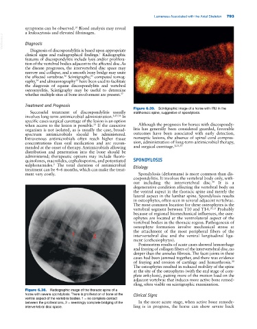
Figure 6.38. Radiographic image of the thoracic spine of a
horse with severe spondylosis. There is proliferation of bone at the Clinical Signs
ventral aspect of the vertebral bodies. 1 = no complete contact
between the proliferations, 2 = seemingly complete bridging of the In the more acute stage, when active bone remode-
intervertebral disc space. ling is in progress, the horse can show severe back