
Page 828 - Adams and Stashak's Lameness in Horses, 7th Edition
P. 828
794 Chapter 6
VetBooks.ir
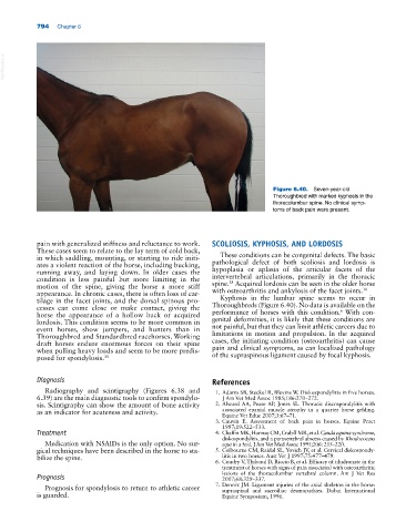
Figure 6.40. Seven‐year‐old
Thorough bred with marked kyphosis in the
thoracolumbar spine. No clinical symp-
toms of back pain were present.
pain with generalized stiffness and reluctance to work. SCOLIOSIS, KYPHOSIS, AND LORDOSIS
These cases seem to relate to the lay term of cold back,
in which saddling, mounting, or starting to ride initi- These conditions can be congenital defects. The basic
ates a violent reaction of the horse, including bucking, pathological defect of both scoliosis and lordosis is
running away, and laying down. In older cases the hypoplasia or aplasia of the articular facets of the
condition is less painful but more limiting in the intervertebral articulations, primarily in the thoracic
38
motion of the spine, giving the horse a more stiff spine. Acquired lordosis can be seen in the older horse
30
appearance. In chronic cases, there is often loss of car- with osteoarthritis and ankylosis of the facet joints.
tilage in the facet joints, and the dorsal spinous pro- Kyphosis in the lumbar spine seems to occur in
cesses can come close or make contact, giving the Thoroughbreds (Figure 6.40). No data is available on the
6
horse the appearance of a hollow back or acquired performance of horses with this condition. With con-
lordosis. This condition seems to be more common in genital deformities, it is likely that these conditions are
event horses, show jumpers, and hunters than in not painful, but that they can limit athletic careers due to
Thoroughbred and Standardbred racehorses. Working limitations in motion and propulsion. In the acquired
draft horses endure enormous forces on their spine cases, the initiating condition (osteoarthritis) can cause
when pulling heavy loads and seem to be more predis- pain and clinical symptoms, as can localized pathology
posed for spondylosis. 30 of the supraspinous ligament caused by focal kyphosis.
Diagnosis References
Radiography and scintigraphy (Figures 6.38 and 1. Adams SB, Steckel R, Blevins W. Diskospondylitis in five horses.
6.39) are the main diagnostic tools to confirm spondylo- J Am Vet Med Assoc 1985;186:270–272.
sis. Scintigraphy can show the amount of bone activity 2. Alward AA, Pease AP, Jones SL. Thoracic discospondylitis with
as an indicator for acuteness and activity. associated epaxial muscle atrophy in a quarter horse gelding.
Equine Vet Educ 2007;3:67–71.
3. Cauvin E. Assessment of back pain in horses. Equine Pract
1997;19:522–533.
Treatment 4. Chaffin MK, Honnas CM, Crabill MR, et al. Cauda equina syndrome,
diskospondylitis, and a paravertebral abscess caused by Rhodococcus
Medication with NSAIDs is the only option. No sur- equi in a foal. J Am Vet Med Assoc 1995;206: 215–220.
gical techniques have been described in the horse to sta- 5. Colbourne CM, Raidal SL, Yovich JV, et al. Cervical diskospondy-
bilize the spine. litis in two horses. Aust Vet J 1997;75:477–479.
6. Coudry V, Thibaud D, Riccio B, et al. Efficacy of tiludronate in the
treatment of horses with signs of pain associated with osteoarthritic
lesions of the thoracolumbar vertebral column. Am J Vet Res
Prognosis 2007;68:329–337.
Prognosis for spondylosis to return to athletic career 7. Denoix JM. Ligament injuries of the axial skeleton in the horse:
supraspinal and sacroiliac desmopathies. Dubai International
is guarded. Equine Symposium, 1996.