
Page 833 - Adams and Stashak's Lameness in Horses, 7th Edition
P. 833
Lameness Associated with the Axial Skeleton 799
RADICULOPATHY
When one or more segmental root nerves in the cervi-
VetBooks.ir cal spine are compressed, damaged, or irritated due to
trauma or enlarged facet joints, lameness symptoms
may occur. In these cases, blocking of the distal limb
5
nerves will give a negative outcome. Physical exam
might show a reduced mobility in the neck, but some-
times the musculoskeletal exam is uneventful. The neu-
rological exam however will show clear signs of nerve
impingement with loss of skin sensitivity in the affected
area, while often the adjacent segments show an augmented
response to skin sensitivity testing. Ultrasonography can
be a useful tool in identifying enlargement of the soft
tissues of the cervical facet joints with enlargement of
the synovial membrane and joint effusion when radio-
graphs are not showing clear indication of affected cer-
vical facet joints. Imaging using Doppler can identify the
most affected region.
Osteoarthritis of facet joints, especially in the more
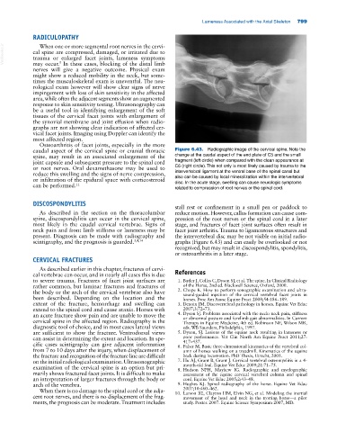
caudal aspect of the cervical spine or cranial thoracic Figure 6.43. Radiographic image of the cervical spine. Note the
spine, may result in an associated enlargement of the change at the caudal aspect of the end plate of C5 and the small
joint capsule and subsequent pressure to the spinal cord fragment (left circle) when compared with the clean appearance at
or root nerves. Oral dexamethasone may be used to C6 (right circle). This not only is most likely caused by trauma to the
reduce this swelling and the signs of nerve compression, intervertebral ligament at the ventral base of the spinal canal but
or infiltration of the epidural space with corticosteroid also can be caused by local mineralization within the intervertebral
disc. In the acute stage, swelling can cause neurologic symptoms
can be performed. 11 related to compression of root nerves or the spinal cord.
DISCOSPONDYLITIS
stall rest or confinement in a small pen or paddock to
As described in the section on the thoracolumbar reduce motion. However, callus formation can cause com-
spine, discospondylitis can occur in the cervical spine, pression of the root nerves or the spinal cord in a later
most likely in the caudal cervical vertebrae. Signs of stage, and fractures of facet joint surfaces often result in
neck pain and front limb stiffness or lameness may be facet joint arthritis. Trauma to ligamentous structures and
present. Diagnosis can be made with radiography and the intervertebral disc may be not visible on initial radio-
scintigraphy, and the prognosis is guarded. 3,4,14 graphs (Figure 6.43) and can easily be overlooked or not
recognized, but may result in discospondylitis, spondylitis,
or osteoarthritis in a later stage.
CERVICAL FRACTURES
As described earlier in this chapter, fractures of cervi-
cal vertebrae can occur, and in nearly all cases this is due References
to severe trauma. Fractures of facet joint surfaces are 1. Butler J, Colles C, Dyson SJ, et al. The spine. In Clinical Radiology
rather common, but laminar fractures and fractures of of the Horse, 2nd ed. Blackwell Science, Oxford, 2000.
the body or the arch of the cervical vertebrae also have 2. Chope K. How to perform sonographic examination and ultra-
been described. Depending on the location and the sound‐guided injection of the cervical vertebral facet joints in
horses. Proc Am Assoc Equine Pract 2008;54:186–189.
extent of the fracture, hemorrhage and swelling can 3. Denoix JM. Discovertebral pathology in horses. Equine Vet Educ
extend to the spinal cord and cause ataxia. Horses with 2007;3:72–73.
an acute fracture show pain and are unable to move the 4. Dyson SJ. Problems associated with the neck: neck pain, stiffness
cervical spine in the affected region. Radiography is the or abnormal posture and forelimb gait abnormalities. In Current
Therapy in Equine Medicine, 4th ed. Robinson NE, Wilson MR,
diagnostic tool of choice, and in most cases lateral views eds. WB Saunders, Philadelphia, 1997.
are sufficient to show the fracture. Ventrodorsal views 5. Dyson, SJ. Lesions of the equine neck resulting in lameness or
can assist in determining the extent and location. In spe- poor performance. Vet Clin North Am Equine Pract 2011;27:
417–437.
cific cases scintigraphy can give adjacent information 6. Faber M. Basic three‐dimensional kinematics of the vertebral col-
from 7 to 10 days after the injury, when displacement of umn of horses walking on a treadmill. Kinematics of the equine
the fracture and recognition of the fracture line are difficult back during locomotion. PhD Thesis, Utrecht, 2001.
on the initial radiological examination. Ultrasonographic 7. Hu AJ, Grant B, Grant J. Cervical vertebral osteomyelitis in a 4‐
month‐old foal. Equine Vet Educ 2009;21:71–75.
examination of the cervical spine is an option but pri- 8. Hudson NPH, Mayhew IG. Radiographic and myelographic
marily shows fractured facet joints. It is difficult to make assessment of the equine cervical vertebral column and spinal
an interpretation of larger fractures through the body or cord. Equine Vet Educ 2005;2:43–48.
arch of the vertebra. 9. Hughes KJ. Spinal radiography of the horse. Equine Vet Educ
When there is no damage to the spinal cord or the adja- 2007;10:460–462.
cent root nerves, and there is no displacement of the frag- 10. Larson BE, Clayton HM, Elvin NG, et al. Modeling the inertial
movement of the head and neck in the trotting horse—a pilot
ments, the prognosis can be moderate. Treatment includes study. Poster. 2007. Equine Science Symposium 2007, MD.