
Page 830 - Adams and Stashak's Lameness in Horses, 7th Edition
P. 830
796 Chapter 6
THE NECK AND POLL
VetBooks.ir Rob Van Wessum
This section emphasizes the cervical disorders that Clinical Signs
directly cause or contribute to lameness. The developmen-
Symptoms of nuchal ligament pathology are diverse
tal cervical disorders, most often causing neurological and very often more visible when the horse is worked.
symptoms, are covered in Chapter 12.
As described in the first part of this chapter, the cervical The horse may appear stiff, the neck does not move up
6
spine has an important function in locomotion. Any and down as much as usual at the walk, and when
dysfunction of the cervical spine can cause an altered worked on circles the head is out with no flexion of the
neck according to the diameter of the circle.
gait pattern, often described as lameness. Sometimes the
When ridden or driven, the contact with the bit
symptoms of the dysfunction of the cervical column are through the reins or lines can feel different to the rider/
observed as altered behavior (head shaking, not con-
necting with the rider’s hand, pulling, bucking, rearing, driver, with one side feeling more rigid or as if the horse
is pulling on one side. Dental issues can have the same
etc.) but are presented to the veterinarian as lameness.
Other cases are presented as lower limb lameness or symptoms, so excluding dental problems helps to con-
firm or at least suspect neck problems. Particular head
shoulder lameness. The following sections cover the
15
common causes of cervical dysfunction and their symp- positions can cause pain, making the horse behave
defensively or reluctant to perform certain exercises.
toms, diagnosis, and treatment.
The neck is an important balancing tool for the horse
during work, so in disciplines in which balance is of
NUCHAL LIGAMENT major importance for performance, such as jumping,
dressage, and eventing, general lack of performance can
The nuchal ligament connects the caudal aspect of be one of the more indistinctive signs.
the skull, dorsal processes of the cervical vertebrae, and
withers region (the dorsal processes of the first thoracic
vertebrae) and has an important function in supporting Diagnosis
the entire neck. Elastic fibers in this structure store Desmopathy can be found at the attachment of the
kinetic energy during locomotion and release the energy ligament to the bone, which may cause enthesophytes
at specific phases of the locomotion when certain muscle that can be seen with radiographs. It also may be located
groups move or activate parts of the cervical spine and in the ligament itself, in which case the altered fiber
the attached front limbs, thus contributing to an effi- pattern is visible on ultrasonographic examination.
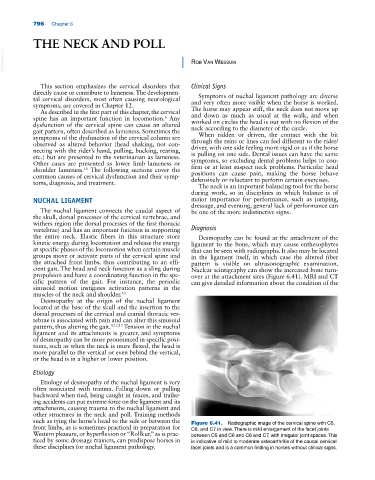
cient gait. The head and neck function as a sling during Nuclear scintigraphy can show the increased bone turn-
propulsion and have a coordinating function in the spe- over at the attachment sites (Figure 6.41). MRI and CT
cific pattern of the gait. For instance, the periodic can give detailed information about the condition of the
sinusoid motion instigates activation patterns in the
muscles of the neck and shoulder. 10
Desmopathy at the origin of the nuchal ligament
located at the base of the skull and the insertion to the
dorsal processes of the cervical and cranial thoracic ver-
tebrae is associated with pain and can alter this sinusoid
pattern, thus altering the gait. 4,12,13 Tension in the nuchal
ligament and its attachments is greater, and symptoms
of desmopathy can be more pronounced in specific posi-
tions, such as when the neck is more flexed, the head is
more parallel to the vertical or even behind the vertical,
or the head is in a higher or lower position.
Etiology
Etiology of desmopathy of the nuchal ligament is very
often associated with trauma. Falling down or pulling
backward when tied, being caught in fences, and trailer-
ing accidents can put extreme force on the ligament and its
attachments, causing trauma to the nuchal ligament and
other structures in the neck and poll. Training methods
such as tying the horse’s head to the side or between the Figure 6.41. Radiographic image of the cervical spine with C5,
front limbs, as is sometimes practiced in preparation for C6, and C7 in view. There is mild enlargement of the facet joints
Western pleasure, or hyperflexion or “Rollkur,” as is prac- between C5 and C6 and C6 and C7, with irregular joint spaces. This
ticed by some dressage trainers, can predispose horses in is indicative of mild to moderate osteoarthritis of the caudal cervical
these disciplines for nuchal ligament pathology. facet joints and is a common finding in horses without clinical signs.