
Page 838 - Adams and Stashak's Lameness in Horses, 7th Edition
P. 838
804 Chapter 7
disruption. It has been shown that in immature cartilage
the deeper layers also possess considerable tensile strength,
G3
VetBooks.ir COOH automatically change with enzymatic degradation of
but this is lost with maturation. The tensile properties
95
hydroxy‐pyridinoline cross‐links, which emphasizes the
importance of these cross‐links in providing cartilage
stiffness, strength, and tension. 95
Collagen fibers are also arranged concentrically
around chondrocytes to form a capsule, called a chon
CS-2 dron. Each chondron contains one or more chondro
Chondroitin cytes, is invested by collagenous pericellular capsule,
sulfate and is surrounded by a proteoglycan‐rich territorial
chains
matrix. Collagen type VI, fibronectin, and thrombos
82
pondin are present in this chondron capsule and help
CS-1 anchor the chondrocyte within the chondron and attach
the chondron within the extracellular matrix. 77
Keratan
sulfate Proteoglycans
chains (KS)
The proteoglycans (previously called mucopolysac
charides) are the other major solid component of the
articular cartilage matrix. They occupy the spaces
between the collagen fibrils. The basic proteoglycan
Link G2 molecule is a monomer formed by a protein core and
protein glycosaminoglycan (GAG) side chains (Figure 7.5).
Most of the proteoglycans (85%) form large aggregates
by noncovalent attachment of the core protein of the
proteoglycan to HA under the stabilization of a link
protein (Figure 7.6). This aggregate is called aggregating
Hyaluronan (HA) G1 (HABR) proteoglycan or aggrecan.
25 nm
The major GAG in adult articular cartilage are
chondroitin‐6‐sulfate and keratan sulfate. Chondroitin‐4‐
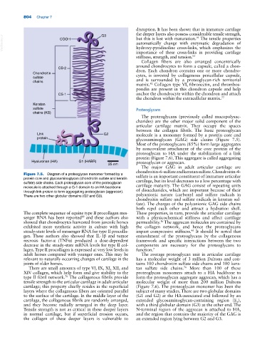
Figure 7.5. Diagram of a proteoglycan monomer formed by a sulfate is an important constituent of immature articular
protein core and glycosaminoglycan (chondroitin sulfate and keratin cartilage, but its level decreases to a low percentage with
sulfate) side chains. Each proteoglycan core of the proteoglycan cartilage maturity. The GAG consist of repeating units
molecule is attached through a G‐1 domain to an HA backbone
through link protein to form aggregating proteoglycan (aggrecan). of disaccharides, which are important because of their
There are two other globular domains (G2 and G3). polyanionic nature (carboxyl and sulfate radicals in
chondroitin sulfate and sulfate radicals in keratan sul
fate). The charges of the polyanionic GAG side chains
both repel each other and attract a hydration shell.
The complete sequence of equine type II procollagen mes These properties, in turn, provide the articular cartilage
senger RNA has been reported and these authors also with a physicochemical stiffness and affect cartilage
88
showed that chondrocytes harvested from juvenile horses permeability. The aggrecan molecules are contained by
56
exhibited more synthetic activity in culture with high the collagen network, and hence the proteoglycans
steady‐state levels of messenger RNA for type II procolla impart compressive stiffness. It should be noted that
46
gen. These authors also showed that IL‐1β and tumor enmeshment of the proteoglycans by the collagenous
necrosis factor‐α (TNFα) produced a dose‐dependent framework and specific interactions between the two
decrease in the steady‐state mRNA levels for type II col components are necessary for the proteoglycans to
lagen. Type II procollagen is expressed at very low levels in function. 7
adult horses compared with younger ones. This may be The average proteoglycan unit in articular cartilage
relevant to naturally occurring changes of cartilage in the has a molecular weight of 3 million Daltons and con
joints of older horses. tains 100 chondroitin sulfate side chains and 100 kera
There are small amounts of type VI, IX, XI, XII, and tan sulfate side chains. More than 100 of these
76
XIV collagen, which help form and give stability to the proteoglycan monomers attach to a HA backbone to
type II fibril network. The collagenous fibrils provide form the proteoglycan aggregate aggrecan, which has a
76
tensile strength to the articular cartilage in adult articular molecular weight of more than 200 million Daltons
cartilage; this property chiefly resides in the superficial (Figure 7.6). The proteoglycan monomer has been the
layers where the collagenous fibers are oriented parallel subject of many studies. There are two globular domains
to the surface of the cartilage. In the middle layer of the (G1 and G2) at the HA‐associated end followed by an
cartilage, the collagenous fibrils are randomly arranged, extended glycosaminoglycan‐containing region (E ),
2
and they become radially arranged in the deep layer. with a third globular domain (G3) at the other end. The
Tensile strength is not as critical in these deeper layers N‐terminal region of the aggrecan is attached to HA,
in normal cartilage, but if superficial erosion occurs, and the region that contains the majority of the GAG is
the collagen of these deeper layers is vulnerable to an extended region lying between G2 and G3.