
Page 836 - Adams and Stashak's Lameness in Horses, 7th Edition
P. 836
802 Chapter 7
VetBooks.ir Collateral ligament
Fibrous joint
capsule
Articular
cartilage Synovial
membrane
Synovial fluid
Subchondral
bone
Figure 7.1. Diagram of a typical synovial joint.
The insertions of the fibrous capsule and articular
ligaments into the adjacent bones demonstrate a zonal
organization with a gradual transition of joint capsule
and ligaments to mineralized fibrocartilage and then to
bone. This enhances the ability of the insertions to dis
tribute forces evenly and decrease the likelihood of pull
out failure. 55
Stability of the joint is provided by the bony configu
ration of the joint, the ligamentous and capsular support
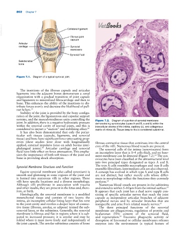
systems, and the musculotendinous units controlling the Figure 7.2. Diagram of a portion of synovial membrane
joint. In addition, there is a negative hydrostatic pressure demonstrating synoviocytes (types A and B, a and b) within the
within the synovial cavity of normal joints, and this is intercellular stroma of the intima, capillary (c), and collagenous
considered to impart a “suction” and stabilizing effect. 85 matrix of intima (d). Tissue deep to this is considered subintimal.
It has also been demonstrated that only the periar
ticular soft tissues (capsule, ligaments, and synovial
tissue) and bone have significant force‐attenuating prop
erties (these studies were done with longitudinally fibrous connective tissue that continues into the central
applied, external impulsive force on adult bovine inter core of the villi. Numerous blood vessels are present.
phalangeal joints). Articular cartilage and synovial The synovial cells of the intima (synoviocytes) form
83
fluid have little effect on force attenuation. This empha an incomplete layer that is 1–4 cells thick, and no base
sizes the importance of both soft tissues of the joint and ment membrane can be detected (Figure 7.2). The syn
63
bone in providing shock absorption. oviocytes have been classified at the ultrastructural level
into two principal types designated as types A and B.
Synovial Membrane Structure and Function The type A cells resemble macrophages and type B cells
resemble fibroblasts. Intermediate cells are also observed.
Equine synovial membrane (also called synovium) is A concept has evolved in which type A and type B cells
smooth and glistening in some regions of the joint and are not distinct, but rather merely cells whose differ
is formed into numerous villi in other regions. These ences in morphology reflect the functions they currently
villi have specific locations and a diverse morphology. perform. 63
Although villi proliferate in association with trauma Numerous blood vessels are present in the subintima
and other insults, they are present in the fetus and, there and extend to within 5–10 μm from the intimal surface. 63
fore, at birth. It appears that each joint has a dual nerve supply con
Histologically, the synovial membrane is a modified sisting of specific articular nerves that reach the joint
mesenchymal tissue and consists of two layers: the capsule as independent articular branches of adjacent
intima, an incomplete cellular lining layer that lies next peripheral nerves and by articular branches that are
to the joint cavity and overlies a deeper layer of connec nonspecific and arise from related muscle nerves. 63
tive tissue (fibrous, areolar, or adipose) termed the sub‐ The three principal functions of the synovial
synovial layer, or the subintima. Generally, the synovial membrane are phagocytosis, regulation of protein and
membrane is fibrous and flat in regions where it is sub hyaluronan (HA) content of the synovial fluid,
jected to increased pressure; it is areolar and may be and regeneration. Excessive phagocytic activity or
63
folded where it must move freely and independently of disruption of lysosomal or cellular membranes releases
the joint capsule. The areolar subintima consists of loose enzymes into the environment (a typical feature of