
Page 837 - Adams and Stashak's Lameness in Horses, 7th Edition
P. 837
Principles of Musculoskeletal Disease 803
VetBooks.ir Articular surface
Tangenital
zone Collagen
fibers
Intermediate
zone
Collagen fibers
(cross section)
Radiate
zone Chondrocytes
Deep zone Calcified cartilage
A B
Subchondral bone
Figure 7.3. Diagram of a metacarpophalangeal joint demon- Tide mark Cancellous bone
strating how redundant synovial membrane gathers at the dorsal
aspect on extension (A) and at the palmar aspect on flexion (B).
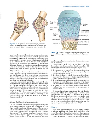
Figure 7.4. Diagram of adult articular cartilage showing the four
layers and the arrangement of the chondrocytes and collagenous
synovitis). The synovial membrane acts as an important fibers.
permeability barrier that, in turn, controls synovial fluid
composition. Most small molecules cross the synovial
membrane by a process of free diffusion that is limited membrane, and periosteum (called the transition zone)
by the intercellular spaces in the synovial membrane and in menisci. 63
101
rather than by blood vessel fenestrations. In traumatic Histologically, adult articular cartilage has been
effusions, changes in protein content and composition divided into four layers, and the chondrocytes have dif
have been associated with both increased vascular ferent appearances within these layers (Figure 7.4):
permeability and increased protein synthesis by the
synoviocytes. 1. The tangential or superficial layer, containing flat
The ability of the synovial membrane to reform fol tened or ovoid chondrocytes and tangentially ori
lowing synovectomy has been described in the horse 40,107 ented collagenous fibrils
and reveals that 120 days after subtotal synovectomy, 2. The intermediate or middle layer, containing larger
there is evidence of restoration and an intimal layer was chondrocytes that may be single or paired and ran
present. However, the synovium was devoid of villi domly oriented collagenous fibrils
107
and there was subintimal fibrosis. 3. The radiate or deeper layer, containing chondrocytes
Another important property of the joint capsule is its arranged in vertical columns separated by collagen
ability to allow complete range of motion. The example ous fibrils that have an overall radial arrangement
of a metacarpophalangeal (fetlock) joint, illustrated in 4. The calcified cartilage layer, composed of mineral
Figure 7.3, shows that synovial membrane gathers at the ized cartilage and chondrocytes in various stages of
dorsal aspect of the joint in extension and at the palmar degeneration
aspect in flexion. This property of gathering is called A basophilic‐staining, undulating line of division
“redundancy.” Inflammation and fibrosis impede this between the radiate layer and the layer of calcified carti
101
property and result in joint stiffness. Elasticity of the lage is termed the “tide mark” or “tide line.” It delineates
joint capsule is also important because of its role in the elastic, nonmineralized layers of the articular carti
shock absorption. 85
lage from the layers of calcified cartilage that has little
resilience. The extracellular matrix of the articular carti
lage is a complex of collagen fibrils, proteoglycans, gly
Articular Cartilage Structure and Function
coproteins, and water (Figures 7.5 and 7.6). 63
Grossly, normal articular cartilage appears milky and
opaque in the thicker regions and translucent with a Collagens
slight bluish tinge in the thinner regions. The surface is
not smooth. Studies using the scanning electron micro Type II collagen comprises 90%–95% of the collagen
scope have demonstrated undulations and irregular in articular cartilage and forms fibrils and fibers inter
depressions. The articular cartilage of equine joints is twined throughout the matrix. Equine type II collagen has
28
generally of the hyaline type, but fibrocartilage is also been characterized biochemically by cyanogen bromide‐
present at the junction of articular cartilage, synovial cleaved peptide profiles and is secreted as a procollagen.
111