
Page 811 - Adams and Stashak's Lameness in Horses, 7th Edition
P. 811
Lameness Associated with the Axial Skeleton 777
SACROILIAC REGION
VetBooks.ir Rob Van Wessum
Diseases of the sacroiliac region are recognized more the joint itself, and a package of small fibrous ligaments
and more as a cause for low‐grade lameness or lack of known as the ventral sacral ligaments. On the top of the
12
performance. 2,3,20 Recent studies show that the sacroiliac pelvic bones, ligamentous structures called the dorsal
joint may not always be involved, but the soft tissue sacral ligaments, originated from the tuber sacrale. These
structures adjacent to the joints in this region can be the include a long ligamentous branch, called the long part,
cause of sacroiliac disease. 19,23,24 that adheres to the fibrous structures of the pelvis (the sac-
The skeletal structures involved in sacroiliac diseases rosciatic ligament), and a shorter portion, called the short
include the pelvic bones and spine, especially the sacro- part, that adheres to the sacral bone and the coccygeal
iliac joints where the ventral aspect of the ilium and ilial vertebrae and ligaments (Figures 6.20–6.22). From the
wing come into close contact with the sacral bone. At ventral aspect of the ilial wing, the interosseus sacroiliac
this junction there are two synovial joints, one at the ligament supports the sacroiliac joint (Figure 6.23). 9,12
ilial facet with a thin layer of fibrocartilage and one at
the sacral facet with hyaline cartilage (Figure 6.19). 9,12,15
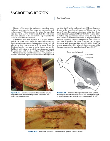
At the ventral aspect of the sacroiliac joint, support is Dorsal sacroilic ligament
provided by several ligamentous structures, the capsule of Short part
Long part
Figure 6.19. Anatomical specimen of the sacroiliac joint, ilial Figure 6.20. Schematic drawing of the dorsal sacral ligament,
wing joint surface, and fibrocartilage. Insert: Sacral bone joint including the short part and long part. Source: Drawing by Maggie
surface and hyaline cartilage. Hoffman. Reprinted with permission from van Wessum , p. 484.
23
Dorsal sacral ligament
Tuber sacrale
Dorsal processes sacral bone
Dorsal sacral ligament (long part)
Sacrosciatic ligament
Figure 6.21. Anatomical specimen of the dorsal sacral ligament, longitudinal view.