
Page 899 - Adams and Stashak's Lameness in Horses, 7th Edition
P. 899
Principles of Musculoskeletal Disease 865
horses with metabolic derangements, however, require Myogenic Atrophy
immediate treatment, including plasma volume expan Muscle atrophy can be myogenic in origin, resulting
VetBooks.ir and cooling (using water and fans). Because most from hypovitaminosis E, severe rhabdomyolysis, immune‐
sion with oral or intravenous isotonic polyionic fluids
11
mediated myositis, disuse, malnutrition, cachexia, and
horses with this condition are alkalotic, administration
of solutions containing sodium bicarbonate is contrain corticosteroid excess. Disuse, malnutrition, cachexia,
and corticosteroid excess are characterized by slow onset
dicated. Daily direct addition of 2 oz. of sodium chloride of atrophy over months of exclusively type 2 fast‐twitch
and 1 oz. of potassium chloride to the feed is recom muscle fibers and normal serum CK. Horses with rhab
mended for horses with recurrent cramping in addition domyolysis and marked elevations in serum CK activ
to electrolyte supplementation before and after endur ity can have a moderate onset of atrophy over a few
ance rides.
weeks. Vitamin E deficiency causes gradual loss of mus
cle mass. 6,16 In contrast, immunemediated myositis
Muscle AtrophyDenervation Atrophy shows rapid loss of 30% of muscle mass over the
Neurogenic atrophy results in fairly rapid atrophy of topline within 48 hours. This disorder has lymphocytic
infiltrates in biopsies of the atrophied muscle associ
both slow‐ and fast‐twitch muscle fibers because the ated with elevated serum CK activity. 18,37
normal low‐level tonic neural stimulus that is necessary
to maintain muscle fiber mass is absent. Complete den
ervation of a muscle results in more than a loss of 50% EXERTIONAL RHABDOMYOLYSIS
of muscle mass within 2–3 weeks. Denervation can be
focal (e.g. suprascapular nerve damage causes supraspi Exertional rhabdomyolysis (ER) occurs in about 3%
natus/infraspinatus atrophy) or generalized with slower of exercising horses and in a wide variety of breeds,
13
progression (e.g. equine motor neuron disease). including draft breeds, Warmbloods, Thoroughbreds,
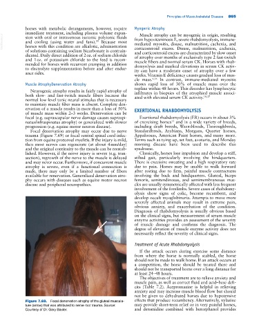
Focal denervation atrophy may occur due to nerve Standardbreds, Arabians, Morgans, Quarter horses,
trauma (Figure 7.69) or focal ventral spinal cord infec Appaloosas, American Paint horses, and many more.
tion from equine protozoal myelitis. If the injury is mild, Terms such as tying‐up, set fast, azoturia, and Monday
then most nerves can regenerate (at about 4 mm/day) morning disease have been used to describe this
and the original continuity to the muscle can be reestab syndrome.
lished. However, if the nerve injury is severe (e.g. tran Classically, horses lose impulsion and develop a stiff,
section), regrowth of the nerve to the muscle is delayed stilted gait, particularly involving the hindquarters.
and may never occur. Furthermore, if concurrent muscle There is excessive sweating and a high respiratory rate
atrophy is severe, even if a functional connection is due to pain. Horses may be unable to walk forward
made, there may only be a limited number of fibers after resting due to firm, painful muscle contractures
available for renervation. Generalized denervation atro involving the back and hindquarters. Gluteal, biceps
phy occurs with diseases such as equine motor neuron femoris, semitendinosus, and semimembranosus mus
disease and peripheral neuropathies. cles are usually symmetrically affected with less frequent
involvement of the forelimbs. Severe cases of rhabdomy
olysis show signs of colic, become recumbent, and
develop occult myoglobinuria. Attempts to move more
severely affected animals may result in extreme pain,
obvious anxiety, and exacerbation of the condition.
Diagnosis of rhabdomyolysis is usually obvious based
on the clinical signs, but measurement of serum muscle
enzyme activities provides an assessment of the severity
of muscle damage and confirms the diagnosis. The
degree of elevation of muscle enzyme activity does not
necessarily reflect the severity of clinical signs.
Treatment of Acute Rhabdomyolysis
If the attack occurs during exercise some distance
from where the horse is normally stabled, the horse
should not be made to walk home. If an attack occurs at
a competition, the horse should be treated there and
should not be transported home over a long distance for
at least 24–48 hours.
The objectives of treatment are to relieve anxiety and
muscle pain, as well as correct fluid and acid–base defi
cits (Table 7.2). Acepromazine is helpful in relieving
anxiety and may increase muscle blood flow but should
not be given to dehydrated horses due to hypotensive
Figure 7.69. Focal denervation atrophy of the gluteal muscula- effects that produce recumbency. Alternatively, xylazine
ture (arrow) that was attributed to nerve root trauma. Source: may provide short‐term relief or in very painful horses,
Courtesy of Dr. Gary Baxter. and detomidine combined with butorphanol provides