Page 1043 - Equine Clinical Medicine, Surgery and Reproduction, 2nd Edition
P. 1043
1018                                       CHAPTER 9


  VetBooks.ir  Clinical presentation                      Prognosis
                                                          The prognosis depends on the severity of the anae-
           Horses may be presented with pale mucous mem-
           branes and may be depressed and weak if anaemia is
           severe. Urine may be red-tinged due to haemoglo-  mia and the presence of secondary organ damage
                                                          from hypoxia. In general, the prognosis is good if
           binuria if there is sufficient intravascular haemolysis.  high levels  of toxins have  not been ingested and
                                                          appropriate supportive care is provided.
           Differential diagnosis
           The differential diagnosis includes red maple  EQUINE INFECTIOUS ANAEMIA
           toxicosis.
                                                          Definition/overview
           Diagnosis                                      EIA, also known as swamp fever, is a viral infection
           A history of onion or garlic ingestion, or administra-  causing disease in all equids including horses, mules,
           tion of phenothiazine, together with laboratory evi-  donkeys and ponies. It occurs worldwide.
           dence supportive of haemolytic anaemia, including
           finding Heinz bodies on the blood smear, confirms  Aetiology/pathophysiology
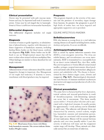
           the diagnosis (Fig. 9.20). Heinz bodies can be dif-  EIA is caused by equine infectious anaemia virus
           ficult to see on Wright-stained smears but will be   (EIAV), a non-oncogenic retrovirus. The virus can
           more evident when stained with new methylene blue.   exhibit latency, and recrudescence can occur inter-
           Other findings are similar to those described for red   mittently. EIAV is transmitted to a susceptible host
           maple toxicosis.                               by an insect vector (tabanid flies, deer flies, stable
                                                          flies and mosquitoes) or fomite (blood-contaminated
           Management                                     needles, syringes, surgical instruments, blood prod-
           Administration of toxic substances should be ceased.   ucts) and enters cells of the mononuclear–phagocytic
           Supportive therapy is important. (See Management   system and endothelial cells. The disease is charac-
           of red maple leaf toxicosis.) If anaemia is severe,   terised by three distinct stages: acute, chronic and
           transfusion with blood products may be required.  inapparent (Fig. 9.21). Haematological abnormali-
                                                          ties exist because of immune-mediated erythrocyte
                                                          destruction and suppressed production of cells in the
                                                          bone marrow.
           9.20
                                                          Clinical presentation
                                                          The acute form is characterised by fever, depression,
                                                          ventral oedema and mucosal petechiation (a result
                                                          of thrombocytopenia). Life-threatening epistaxis
                                                          can occur but is uncommon. This form usually
                                                          occurs within 1–4 weeks after infection and is asso-
                                                          ciated with a high-level viraemia. It usually persists
                                                          for approximately 1 week and may not be identified
                                                          in mild cases. The subacute and chronic forms are
                                                          characterised by anorexia, ventral oedema, weight
                                                          loss, haemolytic anaemia and intermittent pyrexia.
                                                          These signs are associated with recurrent episodes
                                                          of viral replication. Horses will be normal between
                                                          episodes and, over time (usually within 1 year) the
           Fig. 9.20  Blood smear from a horse with Heinz   severity of infection will wane and the inapparent
           body haemolytic anaemia. The arrows indicate Heinz   carrier stage will develop. These animals are clini-
           bodies (Wright’s stain).                       cally normal but are reservoirs of infection.

