Page 1047 - Equine Clinical Medicine, Surgery and Reproduction, 2nd Edition
P. 1047
1022                                       CHAPTER 9


  VetBooks.ir  in adult horses and is most often associated with  Differential diagnosis
                                                          The main differential is AID, which in rare circum-
           chronic external blood loss.
           Aetiology/pathophysiology                      stances can produce microcytic RBCs.
           Chronic external blood loss, which can occur with  Diagnosis
           parasitism, bleeding GI ulcers, masses or coagulopa-  The presence of a microcytic, hypochromic
           thy, is the most common cause. Poor iron intake in   anaemia is almost pathognomonic for iron defi-
           neonates on a milk diet and without access to soil   ciency. Serum iron is decreased and TIBC, which
           may also result in iron-deficiency anaemia.    is essentially a measure of transferrin, is normal
             When iron stores become depleted, developing   to  increased.  Bone  marrow  iron  stores  are  usu-
           RBCs in the bone marrow do not become fully hae-  ally sparse or absent and the G:E ratio may be
           moglobinised and may undergo additional cellular   decreased due to accumulation of the later stages
           division in an attempt to remain in the bone marrow   of developing RBCs that cannot complete matura-
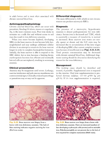
           longer in order to complete haemoglobin formation.   tion because of insufficient iron (Figs. 9.23, 9.24).
           Initially, the bone marrow is able to respond to the   Total protein concentration may be decreased
           RBC deficit, but as iron becomes a limiting factor,   with chronic external blood loss. Additional diag-
           erythropoiesis becomes less efficient and eventually   nostic testing should be directed at identifying the
           lost red cells are not replaced, resulting in worsening   reason for the iron deficiency.
           anaemia.
                                                          Management
           Clinical presentation                          The inciting cause should be identified and
           Anaemia may be inapparent until severe. Lethargy,   treated, if possible. Supplemental iron is required
           exercise intolerance and pale mucous membranes are   in the interim. Oral iron supplementation is pre-
           common initial signs. Clinically evident  haemorrhage   ferred (ferrous sulphate 1.0–4.0 g/450 kg p/o
           or parasitism may or may not be apparent.      q24 h). If i/v iron supplementation is required,


           9.23                                           9.24


           Fig. 9.23  Bone marrow core biopsy from a      Fig. 9.24  Bone marrow core biopsy from a horse with
           horse with iron-deficiency anaemia. The lack of   iron-deficiency anaemia. There are increased numbers of
           blue staining indicates negative staining for iron   late-developing erythrocytic cells (arrows) and no stainable
           (Perl iron stain).                             iron. Polychromatophils are not present due to the lack of
                                                          iron required for complete maturation (H&E stain).

