
Page 158 - Equine Clinical Medicine, Surgery and Reproduction, 2nd Edition
P. 158
Musculoskeletal system: 1.4 The forelimb 133
VetBooks.ir Differential diagnosis 1.235
P1 or distal third metacarpal/metatarsal fracture;
fetlock subluxation; severe fetlock joint injury
(sprain); suspensory branch injury; palmar/plantar
annular ligament injury; digital flexor tendon sheath
pathology; synovial sepsis.
Diagnosis
Clinical examination should alert the clinician to
the possibility of a PSB fracture, although in some
cases lameness can resolve rapidly. Radiography is
important to identify the nature of the fracture and
presence of other damage (e.g. small metacarpal/
metatarsal bone fracture). Multiple views and angles
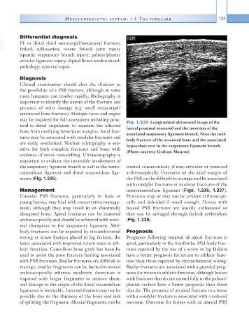
may be required for full assessment including prox- Fig. 1.235 Longitudinal ultrasound image of the
imal-to-distal angulation to separate the affected lateral proximal sesamoid and the insertion of the
bone from overlying bone/joint margins. Axial frac- associated suspensory ligament branch. Note the mid-
tures may be associated with condylar fractures and body fracture of the sesamoid bone and the associated
are easily overlooked. Nuclear scintigraphy is sen- hypoechoic tear in the suspensory ligament branch.
sitive for both complete fractures and bone with (Photo courtesy Graham Munroe)
evidence of stress remodelling. Ultrasonography is
important to evaluate the invariable involvement of
the suspensory ligament branch as well as the inters- treated conservatively if non-articular or removed
esamoidean ligament and distal sesamoidean liga- arthroscopically. Fractures on the axial margin of
ments (Fig. 1.235). the PSB can be difficult to manage and be associated
with condylar fractures or avulsion fractures of the
Management intersesamoidean ligament (Figs. 1.236, 1.237).
Uniaxial PSB fractures, particularly in foals or Fractures may or may not be evident arthroscopi-
young horses, may heal with conservative manage- cally and debrided if small enough. Horses with
ment, although they may result in an abnormally biaxial PSB fractures are usually euthanased or
elongated bone. Apical fractures can be removed they can be salvaged through fetlock arthrodesis
arthroscopically and should be achieved with mini- (Fig. 1.238).
mal disruption to the suspensory ligament. Mid-
body fractures can be repaired by circumferential Prognosis
wiring or screw fixation placed in lag fashion, the Prognosis following removal of apical fractures is
latter associated with improved return rates to ath- good, particularly in the hindlimbs. Mid-body frac-
letic function. Cancellous bone graft has been be tures repaired by the use of a screw in lag fashion
used to assist the poor fracture healing associated have a better prognosis for return to athletic func-
with PSB fractures. Basilar fractures are difficult to tion than those repaired by circumferential wiring.
manage; smaller fragments can be burred/removed Basilar fractures are associated with a guarded prog-
arthroscopically whereas moderate dissection is nosis for return to athletic function, although horses
required with larger fragments to remove them, with fractures that do not extend fully to the palmar/
and damage to the origin of the distal sesamoidean plantar surface have a better prognosis than those
ligaments is inevitable. Internal fixation may not be that do. The presence of an axial fracture in a horse
possible due to the thinness of the bone and risk with a condylar fracture is associated with a reduced
of splitting the fragment. Abaxial fragments can be outcome. Outcome for horses with an abaxial PSB