
Page 240 - Equine Clinical Medicine, Surgery and Reproduction, 2nd Edition
P. 240
Musculoskeletal system: 1.5 The hindlimb 215
VetBooks.ir rarely but is well recognised and possibly hereditary 1.410
in Norwegian Dole ponies. This syndrome is usually
bilateral, involving malformation of the acetabulum
and the head and neck of the femur, which leads to
instability, subluxation and OA, which may also be a
sequela to any of the other conditions affecting the
coxofemoral joint. OCD and OCLLs occur very
rarely in the coxofemoral joint in young horses and
predispose to the early occurrence of OA.
Figs. 1.410, 1.411
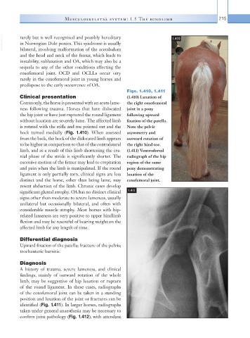
Clinical presentation (1.410) Luxation of
Commonly, the horse is presented with an acute lame- the right coxofemoral
ness following trauma. Horses that have dislocated joint in a pony
the hip joint or have just ruptured the round ligament following upward
without luxation are severely lame. The affected limb fixation of the patella.
is rotated with the stifle and toe pointed out and the Note the pelvic
hock turned medially (Fig. 1.410). When assessed asymmetry and
from the back, the hock of the dislocated limb appears outward rotation of
to be higher in comparison to that of the contralateral the right hind toe.
limb, and as a result of this limb shortening the cra- (1.411) Ventrodorsal
nial phase of the stride is significantly shorter. The radiograph of the hip
excessive motion of the femur may lead to crepitation region of the same
and pain when the limb is manipulated. If the round pony demonstrating
ligament is only partially torn, clinical signs are less luxation of the
distinct and the horse, other than being lame, may coxofemoral joint.
resent abduction of the limb. Chronic cases develop
significant gluteal atrophy. OA has no distinct clinical 1.411
signs other than moderate to severe lameness, usually
unilateral but occasionally bilateral, and often with
considerable muscle atrophy. Most horses with hip-
related lameness are very positive to upper hindlimb
flexion and may be resentful of bearing weight on the
affected limb for any length of time.
Differential diagnosis
Upward fixation of the patella; fracture of the pelvis;
trochanteric bursitis.
Diagnosis
A history of trauma, severe lameness, and clinical
findings, mainly of outward rotation of the whole
limb, may be suggestive of hip luxation or rupture
of the round ligament. In these cases, radiographs
of the coxofemoral joint can be taken in a standing
position and luxation of the joint or fractures can be
identified (Fig. 1.411). In larger horses, radiographs
taken under general anaesthesia may be necessary to
confirm joint pathology (Fig. 1.412), with attendant