
Page 262 - Equine Clinical Medicine, Surgery and Reproduction, 2nd Edition
P. 262
Musculoskeletal system: 1.7a The axial skeleton – neck 237
VetBooks.ir CERVICAL VERTEBRAL MALFORMATION Diagnosis
(SEE ALSO PAGE 1059)
Radiographic examination of the cervical vertebrae
Definition/overview using true laterolateral images permits diagnosis in
some horses. Abnormalities include narrowing of the
Cervical vertebral malformation refers to acquired dorsoventral sagittal diameter of the vertebral canal,
variants in the shape and alignment of the cervical short pedicles, low-slung articular process joints, a
vertebrae, which can result in compression of the wedge-shaped vertebral canal, dorsal enlargement of
cervical spinal cord and ataxia. the caudal epiphysis of a vertebra (ski-jump appear-
ance), extension of the dorsal lamina of a vertebra
Aetiology/pathophysiology and malalignment (subluxation) of adjacent vertebrae
The precise aetiology of cervical vertebral malfor- (Fig. 1.446). Semi-quantitative methods of assess-
mation remains unclear. There is a relationship with ment of vertebral canal dimensions (e.g. intra- and
osteochondrosis and a propensity for rapid growth. intersagittal ratios) have been utilised, but have lim-
There may be a genetic predisposition. ited sensitivity and specificity. Myelography may
assist in determining the site or sites of spinal cord
Clinical presentation compression, but false-negative and false-positive
Horses present with ataxia and weakness of variable results can occur.
severity. The age of onset is highly variable; however,
clinical signs commonly occur in horses 1 to 4 years Management
of age. Affected horses may superficially appear to If clinical signs are observed in young foals, strict
be loose, extravagantly moving horses. Clinical signs dietary management with reduction of energy input
are usually bilaterally symmetrical; the hindlimbs may result in improvement in clinical signs. In care-
are usually more severely affected than the fore- fully selected patients, surgical fusion of vertebrae
limbs. With mild clinical signs the horse may appear at the site of spinal cord compression can result in
croup high in downward transitions from trot to improvement in clinical signs.
walk, have a bouncy gait in transitions and show
occasional circumduction of the outside hindlimb
when turned in small circles. With more severe 1.446
signs the horse may show frequent circumduction
of the hindlimbs, weakness and both forelimb and
hindlimb dysmetria.
Ce 4
Differential diagnosis
Other possible causes of ataxia and weakness
include neuroaxonal dystrophy, extradural haema-
toma, traumatic causes of spinal cord compression,
spinal cord compression associated with osteoar-
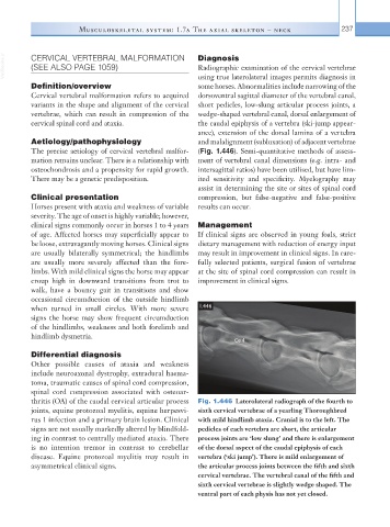
thritis (OA) of the caudal cervical articular process Fig. 1.446 Laterolateral radiograph of the fourth to
joints, equine protozoal myelitis, equine herpesvi- sixth cervical vertebrae of a yearling Thoroughbred
rus 1 infection and a primary brain lesion. Clinical with mild hindlimb ataxia. Cranial is to the left. The
signs are not usually markedly altered by blindfold- pedicles of each vertebra are short, the articular
ing in contrast to centrally mediated ataxia. There process joints are ‘low slung’ and there is enlargement
is no intention tremor in contrast to cerebellar of the dorsal aspect of the caudal epiphysis of each
disease. Equine protozoal myelitis may result in vertebra (‘ski jump’). There is mild enlargement of
asymmetrical clinical signs. the articular process joints between the fifth and sixth
cervical vertebrae. The vertebral canal of the fifth and
sixth cervical vertebrae is slightly wedge shaped. The
ventral part of each physis has not yet closed.