
Page 266 - Equine Clinical Medicine, Surgery and Reproduction, 2nd Edition
P. 266
Musculoskeletal system: 1.7a The axial skeleton – neck 241
VetBooks.ir 1.453 1.454
Ce 4 1
Ce 6
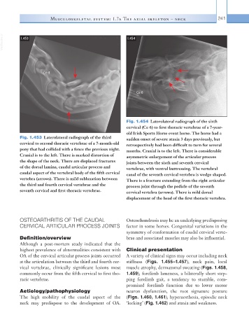
Fig. 1.454 Laterolateral radiograph of the sixth
cervical (Ce 6) to first thoracic vertebrae of a 7-year-
old Irish Sports Horse event horse. The horse had a
Fig. 1.453 Laterolateral radiograph of the third sudden onset of severe ataxia 3 days previously, but
cervical to second thoracic vertebrae of a 7-month-old retrospectively had been difficult to turn for several
pony that had collided with a fence the previous night. months. Cranial is to the left. There is considerable
Cranial is to the left. There is marked distortion of asymmetric enlargement of the articular process
the shape of the neck. There are displaced fractures joints between the sixth and seventh cervical
of the dorsal lamina, caudal articular process and vertebrae, with ventral buttressing. The vertebral
caudal aspect of the vertebral body of the fifth cervical canal of the seventh cervical vertebra is wedge shaped.
vertebra (arrows). There is mild subluxation between There is a fracture extending from the right articular
the third and fourth cervical vertebrae and the process joint through the pedicle of the seventh
seventh cervical and first thoracic vertebrae. cervical vertebra (arrows). There is mild dorsal
displacement of the head of the first thoracic vertebra.
OSTEOARTHRITIS OF THE CAUDAL Osteochondrosis may be an underlying predisposing
CERVICAL ARTICULAR PROCESS JOINTS factor in some horses. Congenital variations in the
symmetry of conformation of caudal cervical verte-
Definition/overview brae and associated muscles may also be influential.
Although a post-mortem study indicated that the
highest prevalence of abnormalities consistent with Clinical presentation
OA of the cervical articular process joints occurred A variety of clinical signs may occur including neck
at the articulation between the third and fourth cer- stiffness (Figs. 1.455–1.457), neck pain, local
vical vertebrae, clinically significant lesions most muscle atrophy, dermatomal sweating (Figs. 1.458,
commonly occur from the fifth cervical to first tho- 1.459), forelimb lameness, a bilaterally short step-
racic vertebrae. ping forelimb gait, a tendency to stumble, com-
promised forelimb function due to lower motor
Aetiology/pathophysiology neuron dysfunction, the root signature posture
The high mobility of the caudal aspect of the (Figs. 1.460, 1.461), hyperaesthesia, episodic neck
neck may predispose to the development of OA. ‘locking’ (Fig. 1.462) and ataxia and weakness.