
Page 270 - Equine Clinical Medicine, Surgery and Reproduction, 2nd Edition
P. 270
Musculoskeletal system: 1.7a The axial skeleton – neck 245
VetBooks.ir 1.463 1.464
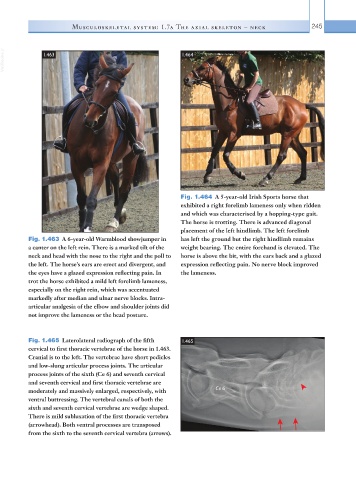
Fig. 1.464 A 5-year-old Irish Sports horse that
exhibited a right forelimb lameness only when ridden
and which was characterised by a hopping-type gait.
The horse is trotting. There is advanced diagonal
placement of the left hindlimb. The left forelimb
Fig. 1.463 A 6-year-old Warmblood showjumper in has left the ground but the right hindlimb remains
a canter on the left rein. There is a marked tilt of the weight bearing. The entire forehand is elevated. The
neck and head with the nose to the right and the poll to horse is above the bit, with the ears back and a glazed
the left. The horse’s ears are erect and divergent, and expression reflecting pain. No nerve block improved
the eyes have a glazed expression reflecting pain. In the lameness.
trot the horse exhibited a mild left forelimb lameness,
especially on the right rein, which was accentuated
markedly after median and ulnar nerve blocks. Intra-
articular analgesia of the elbow and shoulder joints did
not improve the lameness or the head posture.
Fig. 1.465 Laterolateral radiograph of the fifth 1.465
cervical to first thoracic vertebrae of the horse in 1.463.
Cranial is to the left. The vertebrae have short pedicles
and low-slung articular process joints. The articular
process joints of the sixth (Ce 6) and seventh cervical
and seventh cervical and first thoracic vertebrae are
moderately and massively enlarged, respectively, with Ce 6
ventral buttressing. The vertebral canals of both the
sixth and seventh cervical vertebrae are wedge shaped.
There is mild subluxation of the first thoracic vertebra
(arrowhead). Both ventral processes are transposed
from the sixth to the seventh cervical vertebra (arrows).