
Page 376 - Equine Clinical Medicine, Surgery and Reproduction, 2nd Edition
P. 376
Musculoskeletal system: 1.8 Soft-tissue injuries 351
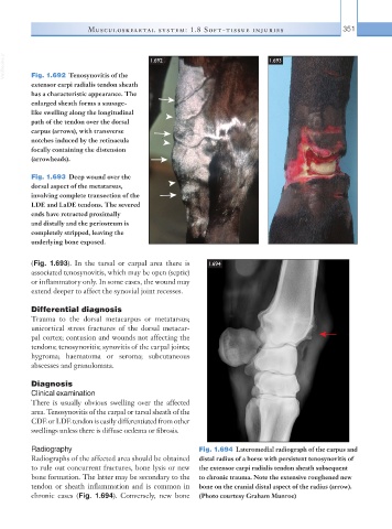
VetBooks.ir Fig. 1.692 Tenosynovitis of the 1.692 1.693
extensor carpi radialis tendon sheath
has a characteristic appearance. The
enlarged sheath forms a sausage-
like swelling along the longitudinal
path of the tendon over the dorsal
carpus (arrows), with transverse
notches induced by the retinacula
focally containing the distension
(arrowheads).
Fig. 1.693 Deep wound over the
dorsal aspect of the metatarsus,
involving complete transection of the
LDE and LaDE tendons. The severed
ends have retracted proximally
and distally and the periosteum is
completely stripped, leaving the
underlying bone exposed.
(Fig. 1.693). In the tarsal or carpal area there is 1.694
associated tenosynovitis, which may be open (septic)
or inflammatory only. In some cases, the wound may
extend deeper to affect the synovial joint recesses.
Differential diagnosis
Trauma to the dorsal metacarpus or metatarsus;
unicortical stress fractures of the dorsal metacar-
pal cortex; contusion and wounds not affecting the
tendons; tenosynovitis; synovitis of the carpal joints;
hygroma; haematoma or seroma; subcutaneous
abscesses and granulomata.
Diagnosis
Clinical examination
There is usually obvious swelling over the affected
area. Tenosynovitis of the carpal or tarsal sheath of the
CDE or LDE tendon is easily differentiated from other
swellings unless there is diffuse oedema or fibrosis.
Radiography Fig. 1.694 Lateromedial radiograph of the carpus and
Radiographs of the affected area should be obtained distal radius of a horse with persistent tenosynovitis of
to rule out concurrent fractures, bone lysis or new the extensor carpi radialis tendon sheath subsequent
bone formation. The latter may be secondary to the to chronic trauma. Note the extensive roughened new
tendon or sheath inflammation and is common in bone on the cranial distal aspect of the radius (arrow).
chronic cases (Fig. 1.694). Conversely, new bone (Photo courtesy Graham Munroe)