
Page 379 - Equine Clinical Medicine, Surgery and Reproduction, 2nd Edition
P. 379
354 CHAPTER 1
VetBooks.ir 1.701 1.702
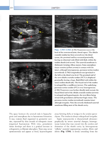
Figs. 1.701–1.703 (1.701) Transverse scan at the
level of the crurotarsal joint, dorsal aspect. The tibialis
cranialis tendon has been severed over the distal
tarsus, the proximal end has retracted proximally,
leaving an abnormal void, filled with fluid, within the
1.703 tendon sheath (red arrow). The synovial membrane is
thickened, forming villous masses. Some amorphous
tissue remains (yellow arrows) in contact with the
intact, although heterogeneous, peroneus tertius tendon
(arrowhead). (1.702) Longitudinal scan (proximal to
the left) at the distal crus level. The proximal end of
the torn tibialis cranialis tendon (TCT) is displaced
proximally, leaving a large, fluid-filled void within the
tarsal tendon sheath (sh). The frayed end of the tendon
is enlarged like a cauliflower (arrow). The underlying
peroneus tertius tendon (PT) is very heterogeneous.
(1.703) Transverse scan further distally (mid-tarsus): the
frayed distal end of the tibialis cranialis tendon (arrows)
is enlarged and hypoechogenic, the torn fibres being
mixed with granulation and haemorrhagic tissue. The
peroneus tertius tendon (arrowhead) remains intact
although irregular. Note the severely thickened synovial
membrane filling most of the sheath cavity.
The space between the severed ends is hypoecho- areas forming clefts or wedges in the tendon paren-
genic and amorphous due to haematoma formation. chyma. The tendon is always enlarged and irregular.
It may contain fluid organised in geometric cavi- Septic tenosynovitis is characterised ultrasono-
ties, separated by thin strands of echogenic tissue graphically by severe synovial changes, heteroge-
(organised haematoma). With time, granulation neous lesions that may extend into the tendon and
tissue develops and there is a gradual increase in distension of the sheath cavity by heterogeneous
echogenicity as fibrosis takes place. Tears may occur ‘cellular’ material representing exudate, fibrin and
spontaneously and appear as focal, hypoechogenic debris (Fig. 1.704). A fistula extending from the