
Page 599 - Equine Clinical Medicine, Surgery and Reproduction, 2nd Edition
P. 599
574 CHAPTER 2
VetBooks.ir 2.170 2.171
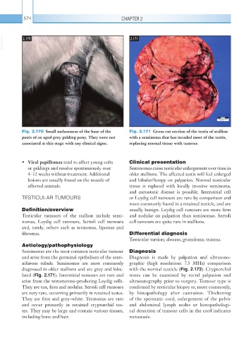
Fig. 2.170 Small melanomas of the base of the Fig. 2.171 Gross cut section of the testis of stallion
penis of an aged grey gelding pony. They were not with a seminoma that has invaded most of the testis,
associated at this stage with any clinical signs. replacing normal tissue with tumour.
• Viral papillomas tend to affect young colts Clinical presentation
or geldings and resolve spontaneously over Seminomas cause testicular enlargement over time in
4–12 weeks without treatment. Additional older stallions. The affected testis will feel enlarged
lesions are usually found on the muzzle of and lobular/lumpy on palpation. Normal testicular
affected animals. tissue is replaced with locally invasive seminoma,
and metastatic disease is possible. Interstitial cell
TESTICULAR TUMOURS or Leydig cell tumours are rare by comparison and
more commonly found in a retained testicle, and are
Definition/overview usually benign. Leydig cell tumours are more firm
Testicular tumours of the stallion include semi- and nodular on palpation than seminomas. Sertoli
nomas, Leydig cell tumours, Sertoli cell tumours cell tumours are quite rare in stallions.
and, rarely, others such as teratomas, lipomas and
fibromas. Differential diagnosis
Testicular torsion; abscess; granuloma; trauma.
Aetiology/pathophysiology
Seminomas are the most common testicular tumour Diagnosis
and arise from the germinal epithelium of the semi- Diagnosis is made by palpation and ultrasono-
niferous tubule. Seminomas are most commonly graphic (high resolution: 7.5 MHz) comparison
diagnosed in older stallions and are grey and lobu- with the normal testicle (Fig. 2.172). Cryptorchid
lated (Fig. 2.171). Interstitial tumours are rare and testes can be examined by rectal palpation and
arise from the testosterone-producing Leydig cells. ultrasonography prior to surgery. Tumour type is
They are tan, firm and nodular. Sertoli cell tumours confirmed by testicular biopsy or, more commonly,
are very rare, occurring primarily in retained testes. by histopathology after castration. Thickening
They are firm and grey–white. Teratomas are rare of the spermatic cord, enlargement of the pelvic
and occur primarily in retained cryptorchid tes- and abdominal lymph nodes or histopathologi-
tes. They may be large and contain various tissues, cal detection of tumour cells in the cord indicates
including bone and hair. metastasis.