
Page 609 - Equine Clinical Medicine, Surgery and Reproduction, 2nd Edition
P. 609
584 CHAPTER 2
VetBooks.ir 2.184 2.185
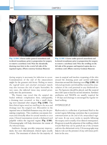
Fig. 2.184 A horse under general anaesthesia and Fig. 2.185 A horse under general anaesthesia and
in dorsal recumbency prior to preparation for surgery in dorsal recumbency prior to preparation for surgery
to remove a scirrhous cord. Note the chronically to remove a scirrhous cord. Note the swelling on the
draining tract hole in the cranial left side of the left side of the prepuce and inguinal region due to a
inguinal region. (Photo courtesy Graham Munroe) scirrhous cord. (Photo courtesy Graham Munroe)
during surgery is necessary for infection to occur. cases is surgical and involves reopening of the skin
Contamination of the end of the emasculators around the draining point and careful soft-tissue
might be the greatest risk factor. Failing to resect dissection around the draining tract (Fig. 2.186). All
the vaginal tunic and external cremaster muscle infected spermatic cord should be removed by emas-
may also increase the risk of septic funiculitis. In culation of the cord proximal to any thickened tis-
rare cases, the infected tissue may extend proxi- sue. No ligatures should be placed, and the surgical
mally into the peritoneum. site is left open to heal by second intention. Systemic
The history may reveal that the surgical site antibiotics and NSAIDs are usually required for
healed without complication then, subsequently, 5–10 days and drainage is encouraged by regular in-
a draining tract occurred, or that a small drain- hand walking.
ing tract remained after surgery (Fig. 2.184). The
first clinical signs noted are swelling in the area and HYDROCOELE
drainage near the surgical scar. Discomfort in the
inguinal area or hindlimb lameness may also be pre- Hydrocoele is a collection of peritoneal fluid in the
senting complaints. This condition can remain dor- scrotal region following castration due to persistent
mant and clinically silent for several months or even communication at the end of the emasculated vagi-
years. Clinical examination reveals a thickened cord nal tunic. It can occur weeks to months following
palpable within the inguinal region (Fig. 2.185). the surgery and is usually of no clinical consequence.
Ultrasonography will help determine the extent of Diagnosis is made by palpation of the swelling, which
the infected tissue. reveals fluid that can be pushed out of the scrotum
Antibiotic therapy provides temporary improve- and into the abdominal cavity. Ultrasonography may
ment, but once discontinued, clinical signs usually help to rule out the presence of any soft-tissue prob-
return. The treatment of choice for the majority of lems in the area.