
Page 637 - Equine Clinical Medicine, Surgery and Reproduction, 2nd Edition
P. 637
612 CHAPTER 3
VetBooks.ir treatment or antiseptic solution treatment, often Diagnosis
Endoscopic examination of the nasal meatus usually
for sinusitis. It is common for the original primary
sinusitis to resolve, but mycotic sinusitis to develop
charge from the nasomaxillary sinus drainage angle.
during treatment. This is a confusing presentation reveals no abnormalities, or possibly scanty dis-
as the clinical signs will often not alter, although Radiography is also frequently unrewarding with
the disease process changes. Mycotic infections can limited signs, possibly some fluid lines in the max-
be secondary to other conditions such as tumours. illary sinuses, but often no abnormalities. Gamma
Primary mycotic sinusitis is recognised, but it is hard scintigraphy frequently reveals an intense increase in
to be definitive that this was the original condition. uptake of the radioisotope within the affected area of
Fungal infections are erosive and in some cases dam- the paranasal sinuses. Care must be taken interpreting
age to the nasal conchae or infraorbital canal can the scan as it is possible to misdiagnose the uptake as a
develop. This can include erosion of the nasomax- periapical tooth abscess. It is important to remember
illary opening, so that an endoscope can be passed that dental disease is not the only cause of increased
directly into the paranasal sinus (Fig. 3.34). uptake of radioisotope in the paranasal sinuses.
Diagnosis requires direct sinus endoscopy
Clinical presentation (sinusoscopy). Direct endoscopy is simply achieved
The clinical presentation is typical of sinusitis. via a trephine hole, either in the caudal maxillary
There is a unilateral nasal discharge and usually or the frontal sinuses. The trephine hole is made in
unilateral enlargement of the submandibular lymph the standing sedated horse under local anaesthesia.
node. There is usually a chronic history and often Orientation of the endoscope within the paranasal
one of previous antibiotic treatment. Facial swell- sinuses can be difficult – the two key features are the
ing is very rare, as is facial ulceration. The discharge sharp-edged frontomaxillary opening and the linear
is frequently malodourous, though less frequently infraorbital canal (Fig. 3.35). Mycotic plaques have
haemorrhagic than nasal mycosis. a typical diphtheritic appearance and microscopic
examination is definitive, with millions of fungal
Differential diagnosis hyphae easily identified (Fig. 3.36).
The differential diagnosis includes almost all other
causes of sinusitis. Due to the chronic nature of the
condition most horses are investigated for secondary 3.35
sinusitis.
3.34
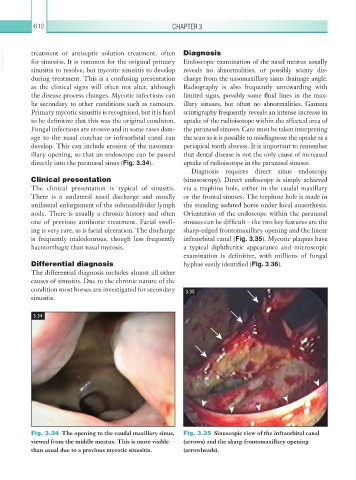
Fig. 3.34 The opening to the caudal maxillary sinus, Fig. 3.35 Sinuscopic view of the infraorbital canal
viewed from the middle meatus. This is more visible (arrows) and the sharp frontomaxillary opening
than usual due to a previous mycotic sinusitis. (arrowheads).